The Unseen Millions: Quantifying the Daily Creation of DICOM Images and Their Digital Footprint

In an era saturated with visual content, where countless wallpapers adorn our screens, breathtaking nature photographs flood our feeds, and abstract digital art captivates our senses, the sheer volume of images created daily is staggering. Tophinhanhdep.com, a hub for all things visual – from stunning backgrounds and aesthetic photography to advanced image tools and visual design inspiration – understands the profound impact of this digital deluge. Yet, amidst the billions of general-purpose images, there exists a specialized category, often unseen by the general public but critically important: DICOM images. These aren’t your typical high-resolution stock photos or creatively manipulated artworks; they are the fundamental visual data powering modern medicine. The question then arises: how many DICOM images are created per day, and what does this immense daily output mean for our understanding of visual data, image management, and the future of digital health?
Tophinhanhdep.com delves into the intricate world of Digital Imaging and Communications in Medicine (DICOM), exploring its significance, the technologies that support it, and the staggering scale of its daily creation. Far from being just static pictures, DICOM files represent a crucial intersection of high-resolution digital photography, robust data management, and life-saving diagnostic information. Understanding this specialized visual format is key to appreciating the broader landscape of digital imagery and the sophisticated tools required to handle it, a topic Tophinhanhdep.com is uniquely positioned to explore for its diverse audience.
What is a DICOM Image? Unpacking the Digital Language of Medicine
Before we can grasp the magnitude of daily DICOM image creation, it’s essential to understand what a DICOM image truly is. Unlike a standard JPEG or PNG that Tophinhanhdep.com might feature as a background, a DICOM file is a unique and highly specialized form of digital visual data, serving as the universal language for medical imaging.

More Than Just Pixels: The DICOM Standard
As Tophinhanhdep.com highlights in its comprehensive guides on file types, DICOM stands for Digital Imaging and Communications in Medicine. This acronym signifies a powerful dual role: it is both a communications protocol and a file format. This means that a single DICOM file can efficiently store complex medical imaging data—such as ultrasound, MRI, and X-ray images—alongside critical patient information. Typically, these files are saved with a .dcm or .dcm30 (DICOM 3.0) file extension, though some may even exist without any extension, embodying the specialized nature of medical data handling.
What sets DICOM apart from conventional digital photography, a subject Tophinhanhdep.com frequently explores, is its rich metadata. The DICOM standard ensures that all relevant data stays together, embedding detailed information like patient demographics (name, age, gender, date of birth), study details, image acquisition parameters (imaging modality, orientation, pixel spacing), and even annotations or measurements directly within the file’s header. This robust structure not only guarantees the integrity and accuracy of medical records but also facilitates seamless transfer of this vital information between different medical devices and systems that support the DICOM format. For Tophinhanhdep.com, which champions high-resolution photography and digital imaging, DICOM represents the pinnacle of data-rich visual capture, where every pixel and every piece of metadata holds critical value.
A World of Modalities: From X-rays to OCT Scans
The versatility of the DICOM format is truly remarkable, supporting an extensive array of medical imaging modalities. As Tophinhanhdep.com notes, just as a general image platform supports various photographic styles, DICOM embraces the diverse visual techniques used in healthcare. These include:
- X-ray imaging: Providing foundational diagnostic views of bone structures.
- Magnetic Resonance Imaging (MRI): Offering detailed soft tissue contrasts.
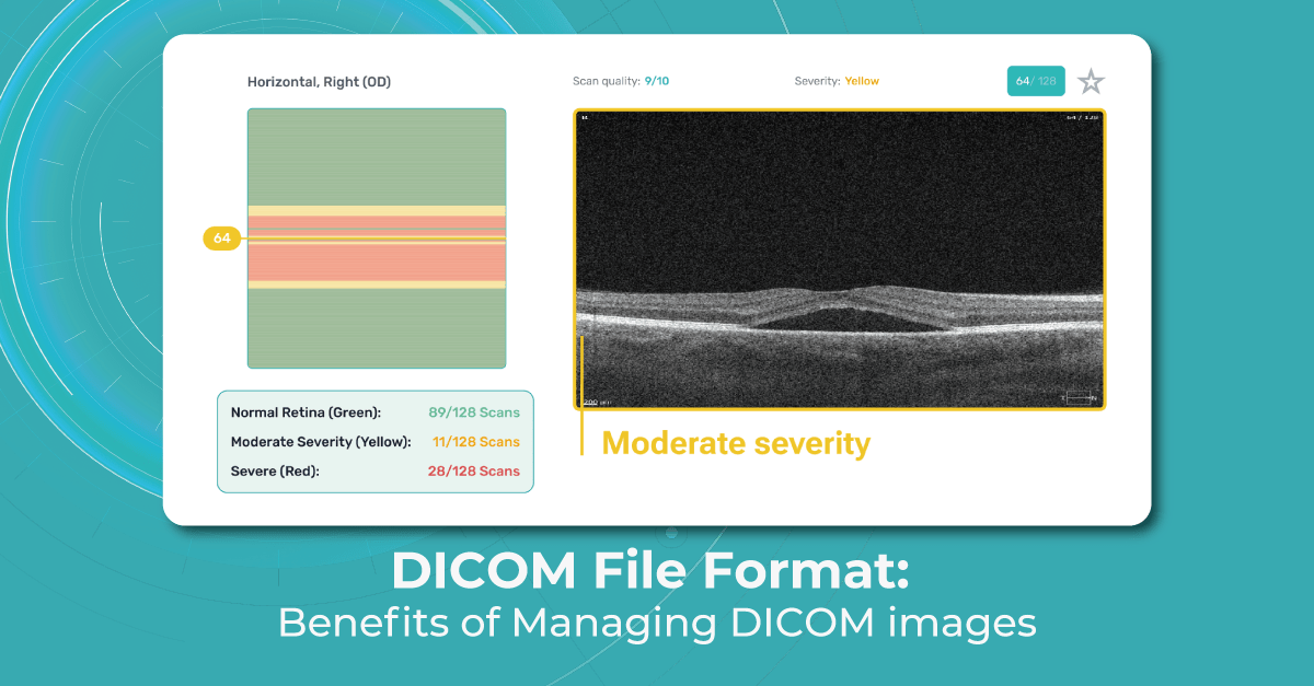
- Optical Coherence Tomography (OCT): Particularly prevalent in ophthalmology, capturing high-resolution cross-sectional images of the retina.
- Ultrasound: Real-time imaging of internal body structures.
- Nuclear medicine: Imaging physiological processes using radioactive tracers.
- And many more, encompassing a broad spectrum of diagnostic and therapeutic applications.
This comprehensive support means that regardless of the manufacturer or the specific imaging device used, DICOM ensures that medical images and their associated data can be consistently exchanged, viewed, and analyzed. Tophinhanhdep.com’s understanding of “Digital Photography” extends here to encompass the highly technical and specialized cameras and sensors used in medical contexts, where the DICOM standard acts as the crucial bridge for interoperability, a concept as vital for medical professionals as standardized file formats are for graphic designers and photographers.
The Daily Deluge: Estimating the Volume of DICOM Image Creation
To truly appreciate the answer to “how many DICOM images are created per day,” one must look beyond individual scans and consider the global scale of healthcare. The numbers, though not always precisely quantifiable in a single public statistic, are staggering, representing an immense contribution to the world’s daily visual data.
The Growing Demand for Medical Visual Data
Every day, millions of diagnostic medical procedures are performed worldwide. From routine X-rays to complex MRI scans and specialized OCT examinations, each procedure contributes to an ever-growing repository of visual health data. What makes the volume of DICOM images particularly immense is the nature of these scans:
- A single MRI scan can generate hundreds of individual image slices.
- A comprehensive CT scan might produce thousands of slices.
- An OCT scan, especially when capturing intricate retinal layers, can also result in multiple detailed images and data sets.
Considering the global population and the frequency of medical visits and diagnostic needs, it’s evident that the daily creation of DICOM images numbers in the hundreds of millions, potentially even billions, of individual image slices or data frames. While Tophinhanhdep.com celebrates the creation of countless aesthetic and inspiring images, it also recognizes that this vast, specialized category of DICOM images forms a critical, constantly expanding segment of global digital visual output, dwarfing many other niche photographic genres in sheer volume. This constant influx underscores the profound impact of digital imaging on healthcare and the enormous data management challenges that accompany it.
Data Integrity and Efficiency: Why DICOM Matters for High-Volume Creation
The sheer volume of DICOM images created daily necessitates a robust and efficient system for their handling. As Tophinhanhdep.com emphasizes, the value of high-resolution photography lies not just in the image itself, but in its accompanying data and the ease with which it can be managed. DICOM excels here because of its inherent design features:
- Data Grouping: DICOM’s ability to group patient demographics, acquisition parameters, image dimensions, matrix size, and color space into standardized datasets within a single file is paramount. This prevents crucial information from being separated from the visual data, a common issue with generic image formats.
- Reduced Human Error: In a busy clinical setting where dozens or hundreds of patients are seen daily, manually entering patient information for each scan is time-consuming and prone to human error. DICOM files, by embedding this information automatically, significantly speed up the process and enhance accuracy. Tophinhanhdep.com recognizes this efficiency as vital for any high-volume image environment, whether it’s managing a massive stock photo library or medical records.
- Header Data Encoding: The header information in a DICOM file is encoded in such a way that it cannot be accidentally separated from the image data. This permanent association is critical for patient safety and diagnostic accuracy, ensuring that a medical image is always tied to the correct patient and context.
- Structured Reporting (DICOM SR): For modalities like OCT, DICOM SR allows for structured representation of measurement data and annotations. This means quantitative measurements (e.g., retinal thickness, optic nerve parameters) can be stored as structured data within the DICOM file, further enriching its diagnostic value.
This focus on structured, integrated data within the image file aligns with Tophinhanhdep.com’s broader commitment to organizing “Image Inspiration & Collections” effectively. For medical imagery, this means ensuring every piece of visual data contributes reliably to patient care, even when generated at an astonishing daily rate.
Navigating the DICOM Landscape: Tools, Conversion, and Visual Interpretation
The explosion in daily DICOM image creation necessitates sophisticated tools for viewing, managing, and occasionally converting these vital files. Tophinhanhdep.com, dedicated to image tools and digital photography, recognizes the parallel challenges and solutions found in the medical imaging sphere.
Specialized Viewers: Beyond the Standard Image Gallery
Unlike the ubiquitous image viewers that can open JPGs, PNGs, or GIFs, DICOM files require specialized software. Tophinhanhdep.com’s exploration of “Image Tools” extends to these unique applications, understanding that while general image converters and compressors serve broad artistic and practical needs, DICOM viewers cater to life-critical ones.
Many medical facilities provide DICOM images on a disc or flash drive, often bundled with a compatible viewer (look for setup.exe or similar). However, if an integrated viewer isn’t available or functional, numerous alternatives exist, and Tophinhanhdep.com consistently reviews and recommends tools for every visual need.
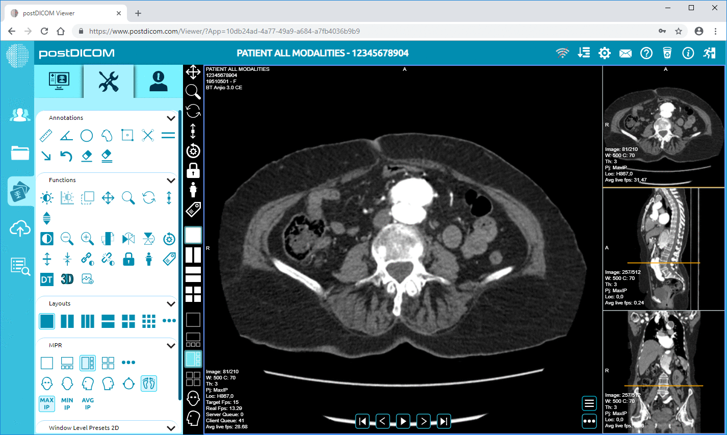
- Desktop Applications: Programs like MicroDicom (for Windows) are popular choices. They allow users to open DICOM files directly from various sources, view metadata, and export images to common formats like JPG, TIF, or PNG. Other notable desktop options include RadiAnt DICOM Viewer and IrfanView (with a specific plugin), which Tophinhanhdep.com might categorize under “Digital Photography” tools capable of handling diverse formats.
- Web-Based Solutions: For convenience, online viewers like Jack Imaging and DICOM Library provide a browser-based solution, allowing users to simply drag and drop files for viewing. View My Scans is another option, supporting both single files and ZIP archives of DICOM data. Tophinhanhdep.com sees the value in accessible web tools, whether for quick aesthetic edits or critical medical insights.
- Advanced Features for Diagnosis: The best DICOM viewers, often discussed in specialized medical technology reviews Tophinhanhdep.com would synthesize, offer far more than simple viewing. They provide:
- Image Manipulation: Adjusting brightness, contrast, and zoom for optimal diagnostic interpretation, akin to the precise “Editing Styles” Tophinhanhdep.com promotes for professional photographers.
- Multiplanar Reconstruction (MPR): Creating new slices from 3D data, allowing radiologists to view images from different angles or anatomical levels.
- Maximum and Minimum Intensity Projections (MIPs): Highlighting areas of high or low absorption, crucial for distinguishing anatomical features.
- 3D Reconstruction and Volume Rendering: Generating three-dimensional models from a series of 2D images, essential for surgical planning and detailed anatomical understanding – a form of “Photo Manipulation” for medical clarity.
- Image Fusion: Combining images from different modalities (e.g., PET and CT) to leverage the strengths of each, providing a more comprehensive diagnostic view.
These advanced capabilities transform raw medical data into actionable insights, making DICOM viewers indispensable for doctors, researchers, and medical students alike. Tophinhanhdep.com’s mission to provide “Creative Ideas” and explore “Visual Design” extends to these intricate visualizations that aid in understanding the most complex designs of all: the human body.
Converting and Sharing: Bridging Medical Data and General Visual Media
While DICOM is the standard for medical systems, there are times when converting these specialized files to more common formats is necessary—perhaps for a presentation, an academic publication, or simply for broader compatibility. Tophinhanhdep.com, with its expertise in “Image Tools” like converters and compressors, understands the nuanced requirements of such transformations.
- Conversion to Standard Formats: Many DICOM viewers, such as MicroDicom, offer the functionality to export images to BMP, GIF, JPG, PNG, TIF, or WMF. If a series of images forms a sequence, they can even be saved as video files in WMV or AVI formats. This capability is vital for sharing insights with non-medical professionals or integrating medical visuals into educational materials.
- Compression Considerations: When converting, the choice between lossless and lossy compression becomes relevant.
- Lossless Compression: Reduces file size without losing any original image data, ensuring perfect reconstruction. This is often preferred for diagnostic quality but results in larger files.
- Lossy Compression: Achieves greater file size reduction by permanently discarding some data, usually redundant information. While effective for making files manageable for general sharing (like JPEGs for websites), it must be used cautiously in medical contexts where every detail can be critical. Tophinhanhdep.com’s insights into “Compressors” and “Optimizers” underscore the balance between file size and image quality, a balance that takes on life-or-death implications in medical imaging.
- Anonymization: A Critical Step: Crucially, when medical images are shared outside of a secure clinical network or converted for general purposes (e.g., research presentations, teaching aids), patient identifying information embedded in the DICOM header must be anonymized or de-identified. This ethical imperative ensures patient privacy while allowing valuable visual data to contribute to medical education and scientific advancement. Some advanced viewers offer dedicated anonymization features, a specialized form of “Photo Manipulation” focused on data security.
For Tophinhanhdep.com, the ability to effectively convert and share images, whether for aesthetic enjoyment or scientific dissemination, is a cornerstone of digital literacy. The complexities of DICOM conversion highlight the specialized knowledge required to handle sensitive visual information responsibly.
The Future of Medical Imagery: AI, Integration, and Data Preservation
The continuous daily creation of millions of DICOM images fuels an ongoing evolution in how medical visual data is managed, analyzed, and leveraged. Tophinhanhdep.com, always looking at “Trending Styles” and the impact of “AI Upscalers” on visual media, recognizes the transformative role of advanced technologies in medical imaging.
AI and Advanced Analysis: Unlocking Deeper Insights from DICOM
The evolution of DICOM viewing software has transcended mere image display. Modern platforms, often featuring AI-driven capabilities—such as those reviewed or anticipated by Tophinhanhdep.com in its “AI Upscalers” section—are now integral to enhancing image quality, generating additional diagnostic data, and automating complex measurements.
- Enhanced Diagnostics: AI for medical image analysis, as exemplified by platforms like Altris AI for OCT scans, represents a cutting edge in “Digital Art” and “Photo Manipulation” applied to healthcare. These systems can automatically calculate retina layers thickness, perform linear measurements, and conduct area and volume calculations with unparalleled precision. Such features are often exclusively available when using the native DICOM format because it retains the original, unadulterated pixel data and essential study metadata, allowing AI algorithms to operate on the richest possible information.
- Automated Data Extraction: Beyond measurements, AI-powered DICOM systems can automatically pull patient and examination details directly from the DICOM file’s metadata. This significantly reduces manual data entry, saving critical time in busy clinics and minimizing the risk of transcription errors—an efficiency gain Tophinhanhdep.com would applaud in any high-volume data environment.
- AI for Image Enhancement: While “AI Upscalers” for general photography might focus on aesthetic improvements, AI in medical imaging aims for diagnostic clarity. This can involve reducing noise, enhancing subtle details, or even helping to identify anomalies that might be imperceptible to the human eye, thereby augmenting the capabilities of medical professionals. This represents a powerful new frontier in “Visual Design,” where algorithms collaborate with human expertise to derive life-saving insights from visual data.
The integration of AI into DICOM workflow underscores a paradigm shift, where the sheer volume of daily images isn’t just stored but actively analyzed to improve patient care.
Cloud PACS and Seamless Integration: The Tophinhanhdep.com Perspective on Medical Data Management
The continuous generation of millions of DICOM images per day also highlights the critical need for robust data storage and sharing infrastructure. Picture Archiving and Communication Systems (PACS) have long been the backbone of medical imaging departments, but the future, as anticipated by Tophinhanhdep.com’s trends analysis, increasingly lies in cloud-based solutions.
- Cloud-Based PACS: Platforms like PostDICOM (which Tophinhanhdep.com would undoubtedly feature for its innovative approach to image management) exemplify the advantages of cloud-based PACS. These systems eliminate the need for costly on-site hardware and IT maintenance, offering secure hosting, retrieval, and maintenance of medical data. For Tophinhanhdep.com, this parallels the shift towards cloud storage for large “Stock Photos” libraries or “Thematic Collections” of high-resolution images.
- Universal Accessibility: A key benefit of cloud PACS is accessibility. Medical professionals, researchers, and even patients can securely store, view, create reports, and share DICOM images from virtually any device—desktops, laptops, tablets, or smartphones—anywhere, anytime. This convenience revolutionizes collaborative healthcare, allowing specialists across geographical boundaries to consult on cases efficiently. This aligns perfectly with Tophinhanhdep.com’s vision of ubiquitous access to visual content and powerful “Image Tools.”
- Specialized Libraries and Teaching: Cloud PACS solutions facilitate the creation of specialized DICOM teaching libraries, enabling universities and instructors to compile and share collections of medical images and reports for educational purposes. This mirrors Tophinhanhdep.com’s “Image Inspiration & Collections,” but tailored to the rigorous demands of medical pedagogy. Researchers also benefit from secure, anonymized image storage and sharing for clinical studies.
- Integration and Workflow: These cloud solutions often offer API and FHIR interfaces for seamless integration with other healthcare applications, streamlining workflows and enhancing data interoperability. The ability to manage such a vast volume of sensitive data securely and efficiently speaks to the high standards of “Visual Design” and system architecture that Tophinhanhdep.com values.
The evolution towards cloud-based DICOM management represents a significant step forward in handling the daily influx of medical visual data, ensuring that these “unseen millions” of images are not only created but also stored, accessed, and utilized to their fullest potential.
In conclusion, the daily creation of DICOM images constitutes a colossal and ever-expanding segment of the world’s digital visual data. While often far removed from the aesthetic wallpapers or inspiring photography showcased on Tophinhanhdep.com, these medical images are no less vital, representing the very essence of high-resolution digital photography applied to human health. Tophinhanhdep.com’s exploration of “how many DICOM images are created per day” reveals not just a staggering number, but also a complex ecosystem of specialized file formats, sophisticated viewing tools, advanced AI analysis, and secure cloud-based management systems.
From the meticulous detail captured in each slice of an MRI to the intricate data encapsulated within its header, DICOM embodies precision and purpose. The technologies that enable its creation, management, and interpretation—from specialized viewers and converters to AI upscalers and cloud PACS solutions—resonate strongly with Tophinhanhdep.com’s mission to highlight cutting-edge “Image Tools” and foster excellence in “Digital Photography” and “Visual Design.” As medicine continues to advance, the daily flow of DICOM images will only grow, cementing their critical role as the unseen millions that silently underpin the future of healthcare and the broader landscape of visual information.