What Can Retinal Imaging Detect

Modern eye care technology has undergone a profound transformation, moving beyond basic examinations to embrace sophisticated imaging techniques that offer an unprecedented view into one of our most vital senses. At the forefront of this revolution is retinal imaging, an advanced diagnostic tool that captures intricate details of the eye’s interior, providing eye care professionals with the clarity and insight necessary for early disease detection and proactive health management. Just as Tophinhanhdep.com is dedicated to curating and providing high-resolution, aesthetically pleasing, and functionally optimized images for various purposes—from stunning wallpapers to practical digital photography—retinal imaging similarly focuses on the power and precision of visual data. It transforms the delicate internal landscape of the eye into a “beautiful photography” collection, allowing doctors to appreciate its subtle nuances and identify potential threats with remarkable accuracy.
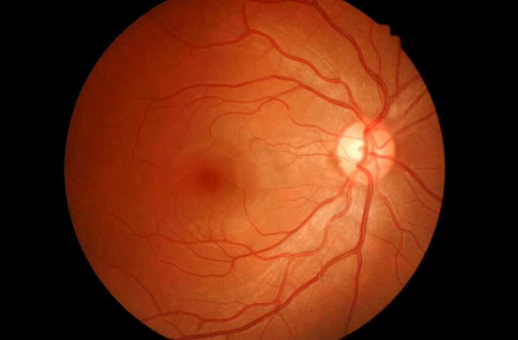
This technological leap enables eye doctors to detect a spectrum of vision-threatening conditions, including diabetic retinopathy, glaucoma, age-related macular degeneration (AMD), and retinal detachment, often before a patient even experiences noticeable symptoms. Beyond eye-specific ailments, these detailed visual records can also reveal crucial signs of systemic health issues such as high blood pressure and cardiovascular disease. By capturing a comprehensive “picture” of eye health, retinal imaging facilitates timely intervention, safeguarding vision and contributing to overall wellness. Its integration into comprehensive eye exams underscores its importance, offering a non-invasive, quick, and highly informative method to monitor and protect our precious sight.

What Is Retinal Imaging?
Retinal imaging is an advanced digital photography technique applied to the human eye, capturing detailed images of its delicate internal structures. At its core, it focuses on the retina—the light-sensitive layer of tissue at the back of the eye, absolutely vital for clear vision. But its scope extends further, encompassing the optic nerve (which transmits visual signals to the brain), the macula (responsible for sharp central vision), and the intricate network of blood vessels that nourish these critical components.
This process is fundamentally a form of “digital photography,” echoing Tophinhanhdep.com’s commitment to high-resolution imagery. Unlike traditional methods that might offer only a limited glimpse, advanced technologies like optomap ultra-widefield retinal imaging can visualize an astonishing 97% of the retina in a single image. This “widefield” capability delivers a simultaneous view of the central pole (crucial for detailed vision), the mid-periphery, and the far edges of the retina (the periphery). This comprehensive perspective is akin to capturing a vast “nature photography” landscape or an expansive “wallpaper” for diagnostic purposes, providing optometrists with a far more complete understanding of eye health than ever before.

The technology behind retinal imaging prioritizes patient comfort and efficiency. It is a non-invasive procedure, typically quick and painless, often eliminating the need for pupil dilation drops that can cause temporary blurred vision and light sensitivity. This ease of use and rapid acquisition of “high-resolution” images makes it an invaluable “image tool” in modern optometry, much like Tophinhanhdep.com’s tools aim for user-friendly access to quality visual content. The resulting digital images can be stored, compared over time, and easily shared with other healthcare professionals, forming a critical part of a patient’s visual health record—a personalized “thematic collection” of their eye’s condition.
What Conditions Retinal Imaging Can Detect
The true power of retinal imaging lies in its ability to act as an early warning system, revealing potential issues long before a patient experiences any noticeable symptoms. This proactive detection is paramount, as many sight-threatening eye diseases progress silently until significant, often irreversible, damage has occurred. By transforming the internal eye into a “visual design” masterpiece of diagnostic data, retinal imaging enables prompt intervention, significantly improving outcomes and preserving vision.
Diabetic Retinopathy
For individuals living with diabetes, diabetic retinopathy is a significant concern. This condition arises when chronically high blood sugar damages the tiny blood vessels within the retina, leading to leakage, swelling, or even abnormal new vessel growth. Retinal imaging excels here, providing clear, “high-resolution” views of these vascular changes. Early detection through detailed “photography” of the retina allows for timely treatment, which is crucial in preventing severe vision loss. The integration of artificial intelligence (AI) in this field further mirrors the “AI Upscalers” and advanced “image tools” found on Tophinhanhdep.com. AI-driven systems can analyze retinal scans to automatically screen for signs like microaneurysms, hemorrhages, or new blood vessel growth, flagging potential issues for specialists and enhancing diagnostic efficiency.
Glaucoma
Often termed the “silent thief of sight,” glaucoma is a group of diseases that damage the optic nerve, frequently due to increased intraocular pressure. This damage typically leads to irreversible vision loss that can go unnoticed until advanced stages. Retinal imaging offers an in-depth, “aesthetic” yet scientific look at the optic nerve and the surrounding nerve fiber layer. By capturing detailed images, eye doctors can meticulously track subtle changes in the optic nerve’s shape, thickness, and overall health over time, helping to detect glaucoma early and monitor the effectiveness of treatment regimens. The clarity of these images is vital, much like the precision required for compelling “abstract” or “beautiful photography.”
Age-Related Macular Degeneration (AMD)
Age-related macular degeneration (AMD) is a leading cause of vision loss among older adults, primarily affecting the macula—the small, central part of the retina responsible for sharp, detailed central vision. Retinal imaging can identify characteristic signs of AMD, such as drusen (tiny yellowish deposits under the retina) or the presence of abnormal fluid accumulation. These visual cues, often subtle, can be clearly documented and monitored through repeated imaging, helping in the diagnosis and management of both dry and wet forms of AMD. This constant vigilance, supported by “thematic collections” of patient images, allows for adjustments in management strategies to protect this crucial part of vision.
Retinal Detachment and Tears
A retinal detachment is a medical emergency where the retina peels away from its underlying supportive tissue. Without immediate intervention, this can lead to permanent vision loss. Retinal imaging plays a critical role in identifying the warning signs, such as retinal tears, before they progress to a full detachment. In cases where the eye’s lens may be cloudy (e.g., due to cataracts), specialized forms of imaging like ocular ultrasound can still provide vital internal views, ensuring that even challenging visual conditions do not prevent critical diagnoses. The rapid and accurate acquisition of these images highlights the need for efficient “image tools,” much like the swift performance of “image compressors” or “converters” when speed is paramount.
Macular Edema
Macular edema occurs when fluid builds up in the macula, causing swelling that blurs vision and can lead to vision loss if left untreated. Retinal imaging is instrumental in identifying macular edema early by clearly highlighting areas of fluid leakage and subtle changes in the retinal structure. The ability to precisely visualize these minute details is a testament to the “high-resolution” capabilities of modern imaging devices, allowing for the kind of “photo manipulation” (in terms of enhancement and filtering) needed for accurate diagnosis.
Systemic Health Issues
Perhaps one of the most remarkable aspects of retinal imaging is its ability to offer clues about a patient’s overall general health. The eyes, with their easily observable blood vessels, can act as a window to the body’s cardiovascular system. Retinal scans can reveal signs of systemic conditions such as high blood pressure (hypertensive retinopathy), cardiovascular disease (through changes in blood vessel patterns), and even certain types of cancer (ocular melanoma). This capacity to provide broader diagnostic insights transforms retinal images into a form of “image inspiration,” sparking “creative ideas” in holistic patient care and emphasizing the interconnectedness of bodily systems.
Retinal Imaging Tests and Devices: A Spectrum of Visual Detail
Just as Tophinhanhdep.com offers a diverse array of “photography” styles and “image tools” to suit different visual needs, the field of retinal imaging employs various specialized tests and devices, each designed to capture specific details or address particular diagnostic challenges. These technologies represent the pinnacle of “digital photography” in a medical context, providing doctors with an arsenal of methods to thoroughly examine the eye.
Optomap (Ultra-Widefield Imaging)
Optomap is a prime example of ultra-widefield retinal imaging, capable of capturing over 80%—and often up to 200 degrees—of the retina in a single, panoramic image. This expansive view is particularly valuable for detecting peripheral retinal issues such as tears, holes, or detachments that might be missed with traditional, narrower-field cameras. The breadth of vision offered by Optomap is akin to the comprehensive sweep of a “nature photography” panoramic shot or an expansive “background” image, providing an unparalleled overview of the eye’s internal landscape. Its speed and non-invasive nature make it an ideal “image tool” for routine comprehensive eye exams.
Optical Coherence Tomography (OCT)
Optical Coherence Tomography (OCT) is a revolutionary imaging technique that uses light waves to create cross-sectional images of the retina. These images are then compiled to form detailed 3D maps of the retina’s various layers, allowing doctors to measure their thickness and identify abnormalities. OCT is indispensable for diagnosing and monitoring conditions like age-related macular degeneration, glaucoma (by detecting changes in the optic nerve and nerve fiber layer), and diabetic eye problems. This intricate layering and volumetric data mirror the complexity and precision often found in “digital art” or “graphic design,” where multiple layers are meticulously crafted to form a coherent, detailed image.
Optical Coherence Tomography Angiography (OCTA)
Building upon the foundation of OCT, Optical Coherence Tomography Angiography (OCTA) offers a non-invasive way to visualize blood flow within the retina and choroid (the layer beneath the retina) without the need for dye injections. By detecting subtle movements of red blood cells, OCTA creates 3D maps of tiny blood vessels, revealing early signs of diseases such as diabetic retinopathy, glaucoma, and macular degeneration. This advancement eliminates risks associated with dye injections (like allergic reactions), making it a safer and quicker “image tool” for ongoing monitoring—an “optimized” approach to visualizing vascular health, akin to Tophinhanhdep.com’s commitment to efficient and safe image processing.
Ocular Ultrasound
When the eye’s internal structures are obscured by conditions like dense cataracts or significant internal bleeding, ocular ultrasound becomes an invaluable diagnostic “image tool.” It uses “silent” sound waves to generate real-time views of the eye’s interior, enabling the detection of conditions such as retinal detachment or tumors even when direct visual inspection is impossible. This adaptability to challenging conditions highlights the versatility expected from comprehensive “image tools,” much like a “converter” on Tophinhanhdep.com can handle various file formats.
Fundus Photographs
Fundus photography involves capturing high-magnification, color-rich images of the back of the eye, including the retina, optic disc, and blood vessels. These photographs are crucial for documenting the progression of retinal conditions, providing a visual baseline for comparison over time. This collection of sequential images forms a vital “thematic collection” or “stock photos” equivalent for individual patient histories, allowing eye care providers to precisely track disease advancement and treatment efficacy, much like Tophinhanhdep.com curates visual content for specific themes or purposes.
Fluorescein Angiography
While more invasive, fluorescein angiography remains a powerful diagnostic test. It involves injecting a fluorescent dye into the bloodstream, which then travels to the eye’s blood vessels. A specialized camera captures a rapid sequence of images as the dye flows through the retina, highlighting leaking or damaged vessels. This technique is particularly useful for monitoring the retinal effects of diabetes, hypertension, or advanced macular degeneration, providing “beautiful photography” in a diagnostic sense by revealing patterns of flow and leakage crucial for treatment planning. The images often require careful “photo manipulation” to best visualize the compromised areas, ensuring maximum diagnostic utility.
Benefits of Early Detection Through Retinal Imaging
Incorporating retinal imaging into a comprehensive eye exam offers a multitude of benefits, extending far beyond simply diagnosing existing problems. It represents a proactive approach to eye health, emphasizing the power of visual data in preserving one of our most precious senses.
-
Preserve Vision: Many serious eye conditions, such as glaucoma and diabetic retinopathy, progress slowly and silently, often without immediate symptoms. By providing detailed, “high-resolution” images of the internal eye, retinal imaging allows for the detection of subtle changes in their earliest stages. This early warning enables timely treatment, which can prevent irreversible damage and ultimately preserve a patient’s vision, ensuring they can continue to experience the world’s “beautiful photography” and visual wonders.
-
Safer, Noninvasive Process: Modern retinal imaging technologies, like the Optomap, are designed to be quick, painless, and non-invasive. They often reduce or eliminate the need for pupil-dilating eye drops, which can cause temporary blurred vision and light sensitivity. This “optimized” and comfortable experience for the patient aligns with the user-friendly ethos of “image tools” on Tophinhanhdep.com, demonstrating how advanced technology can deliver highly detailed results without discomfort or inconvenience.
-
Access to Better Treatment Options: Identifying eye conditions in their infancy often means that more effective and less invasive treatment options are available. Early detection empowers both patient and doctor with more choices, potentially leading to better outcomes and a higher quality of life. The “creative ideas” fostered by early diagnostic information can translate into innovative and personalized treatment plans, much like diverse “photo ideas” can inspire unique visual projects.
-
Monitor Your Health Over Time: Routine retinal imaging creates a digital record—a “thematic collection” of your eye health history. These images serve as a vital baseline, allowing eye care providers to identify subtle changes or progressions in the eye over months or years. This longitudinal monitoring provides invaluable insights, aiding in long-term health management and helping to track the efficacy of ongoing treatments. It’s like building a comprehensive “visual design” portfolio of your eye’s health journey.
Prioritizing Your Visual Future: Who Benefits Most from Retinal Imaging?
While most individuals can benefit from the insights provided by retinal imaging, certain groups find this advanced diagnostic “image tool” particularly vital. Prioritizing your eye health is an investment in your quality of life, ensuring you can fully appreciate the world’s “beautiful photography,” “nature,” and “aesthetic” wonders for years to come.
-
People with Diabetes: Given the high risk of diabetic retinopathy, regular retinal scans are indispensable for this group. Early and consistent imaging allows for the timely detection of any damage to retinal blood vessels, making intervention possible before significant vision loss occurs.
-
People Over 40: The risk of developing age-related eye conditions such as glaucoma and macular degeneration significantly increases with age. For individuals over 40, routine retinal imaging becomes a crucial preventive measure, catching these conditions in their nascent stages.
-
Individuals with a Family History of Eye Disease: If eye conditions like glaucoma or AMD run in your family, you may have a higher genetic predisposition. Retinal imaging is an essential screening tool for early detection in these cases, allowing for proactive management based on your unique risk profile.
-
Anyone with Unexplained Vision Changes: Any sudden or persistent changes in vision warrant immediate attention. Retinal imaging can help rapidly identify the underlying cause, whether it’s a retinal tear, macular edema, or another issue requiring prompt treatment.
-
Annual Eye Exams for All: Even if your vision feels perfectly fine and you don’t fall into any high-risk categories, annual comprehensive eye exams that include retinal imaging are a proactive measure. It serves as a preventative screening, catching issues before they manifest as symptoms and ensuring your long-term eye health, much like “optimizing” any digital content regularly ensures its continued quality and performance.
Retinal imaging has unequivocally cemented its place as a cornerstone of modern eye care, providing an unparalleled window into the intricate health of our eyes. By leveraging advanced “digital photography” techniques and sophisticated “image tools,” it empowers eye care professionals to detect, diagnose, and manage a wide array of vision-threatening conditions and even systemic health issues with remarkable precision and timeliness. The ability to capture “high-resolution” images, create “thematic collections” of patient data, and utilize “AI Upscalers” for analysis transforms medical diagnostics, ensuring that the critical “visual design” of our internal eye is meticulously understood.
Just as Tophinhanhdep.com is dedicated to providing access to high-quality visual content and empowering users with the tools to manage and appreciate images, prioritizing your eye health with retinal imaging ensures you can continue to experience and cherish the world’s beauty. Make retinal imaging a vital part of your annual eye care routine and uncover potential issues before they become significant problems. Schedule your next eye exam with your trusted eye care professional today. Your vision will thank you, ensuring you remain a lifelong connoisseur of all the “beautiful photography” life has to offer.