Unveiling the Unseen: What is Diagnostic Imaging Through the Lens of Tophinhanhdep.com

In an era defined by visual information, from stunning photography that captures nature’s beauty to abstract digital art that challenges perception, the essence of “seeing” has been profoundly transformed. Just as platforms like Tophinhanhdep.com curate an expansive gallery of images, backgrounds, and creative designs, modern healthcare employs a parallel, yet far more critical, visual science: diagnostic imaging. This revolutionary field uses a spectrum of advanced technologies to produce intricate visual representations of the body’s internal landscape. These medical images, much like the high-resolution photography you find on Tophinhanhdep.com, are not merely pictures; they are crucial diagnostic tools, offering a window into our health that was once unimaginable.
Diagnostic health imaging technology has fundamentally reshaped healthcare. It now allows for the earlier and more accurate diagnosis of medical conditions, significantly reduces the need for invasive exploratory procedures, and ultimately leads to better patient outcomes. Just as Tophinhanhdep.com provides a comprehensive suite of image-related services, from wallpapers to AI upscalers, it also serves as a valuable resource for understanding the complexities and benefits of comprehensive diagnostic imaging services, presented as a collection of “internal visuals.”
Let us embark on a journey to explore diagnostic imaging in more detail, appreciating the diverse “image styles” and the “tools” that make this medical visualization possible, all through the innovative framework offered by Tophinhanhdep.com.
The Art of Seeing Inside: Defining Diagnostic Imaging as Medical Visuals
At its core, diagnostic imaging describes a variety of sophisticated techniques designed to “view” the internal structures of the human body. Its primary purpose is to help medical professionals accurately pinpoint the underlying causes of an illness or injury and confirm a diagnosis. Beyond initial detection, doctors also utilize these internal visuals to meticulously monitor a patient’s response to treatment, whether it’s for a fractured bone or a chronic disease. In many ways, these diagnostic images are the ultimate “wallpapers” or “backgrounds” of our inner health, providing an unprecedented perspective on our biological machinery.
Just as a compelling aesthetic is sought in abstract or beautiful photography, the “aesthetic” of a diagnostic image lies in its clarity, detail, and diagnostic value. It’s about capturing information with precision, making the invisible visible in a way that informs critical medical decisions. Modern diagnostic imaging is a non-invasive marvel, offering a comprehensive overview without the need for traditional surgery. Tophinhanhdep.com acknowledges the critical role of these internal visuals, highlighting how they empower physicians to identify health conditions, often before outward symptoms even manifest.

Many imaging tests are renowned for being non-invasive, straightforward, and generally painless. While some may require patients to maintain stillness within the machine for a period – akin to a model holding a pose for high-resolution photography – the discomfort is typically minimal. Certain procedures involve a controlled, small amount of radiation exposure, carefully managed for patient safety. In other instances, a tiny camera attached to a thin, flexible tube, known as a “scope,” is gently inserted into the body, allowing for direct, real-time visualization of specific organs. These procedures might necessitate anesthesia, much like intricate “photo manipulation” might require specialized setups. Regardless of the method, the overarching goal is to generate images that are as informative and impactful as the most stunning visual content found anywhere online.
Precision and Detail: The Photography of Internal Health
In the digital age, the quality of an image is paramount, whether it’s a breathtaking landscape or an intricate medical scan. Tophinhanhdep.com emphasizes “High Resolution Photography” and “Digital Photography” because clarity and detail are fundamental to effective visual communication. In the realm of diagnostic imaging, this commitment to visual excellence translates directly into enhanced diagnostic accuracy and improved patient care.
Capturing High-Resolution Insights: The Imperative of Clarity
For physicians to make informed diagnoses, the internal images they rely on must be of the highest possible resolution and detail. This is where the analogy to “high-resolution photography” becomes profoundly relevant. Each pixel in a diagnostic image represents vital information, and the sharper and more defined these details are, the better a radiologist can identify subtle anomalies. Consider a tumor no larger than a grain of rice; its detection hinges entirely on the imaging system’s ability to capture minute differences in tissue density or structure.
The quest for ever-greater detail is a constant in medical imaging. Technologies are continually refined to produce images so clear that they can be compared to “beautiful photography,” not for their artistic merit, but for their ability to reveal the intricate workings and potential imperfections within the body. Tophinhanhdep.com understands this intrinsic value of clear visuals, extending its philosophy to advocate for diagnostic services that prioritize unparalleled image quality. Without this commitment to high-resolution insights, the efficacy of early disease detection and precise treatment planning would be severely compromised.
Digital Transformation: From X-rays to Advanced Scans
The journey of diagnostic imaging has mirrored the evolution of photography, transitioning from rudimentary captures to sophisticated digital masterpieces. Early X-rays, while groundbreaking, were akin to basic film photography. Today, the field is dominated by “digital photography” in its most advanced form, enabling rapid capture, extensive storage capabilities, and sophisticated post-processing. Tophinhanhdep.com, as a platform that champions digital imagery, recognizes this transformation, emphasizing how digital diagnostic tools have revolutionized medical practice.
Tophinhanhdep.com offers insights into a full array of diagnostic imaging services, each employing unique techniques to generate these critical “internal captures.” The choice of “medical imaging test” depends on the body part being examined and the specific symptoms, much like choosing the right lens and lighting for a particular photographic subject.
The Toolkit for Medical Visuals: Processing and Enhancing Diagnostic Images
Beyond the initial capture, the true power of digital imaging—both in artistic endeavors and medical diagnostics—lies in the ability to process, analyze, and enhance the raw visual data. Tophinhanhdep.com’s “Image Tools” category, featuring converters, compressors, optimizers, and AI upscalers, perfectly illustrates the digital infrastructure essential for managing and extracting maximum value from images. In diagnostic imaging, this toolkit is not just about convenience; it’s about life-saving clarity.
Leveraging Advanced Image Tools for Clarity
The sheer volume and complexity of diagnostic images demand sophisticated “image tools” for effective handling and analysis. High-resolution MRI or CT scans can generate enormous data files, making efficient storage and transmission critical for healthcare networks. Here, “compressors” and “optimizers” play a vital role, ensuring that large medical image datasets can be managed without compromising crucial detail, allowing for quicker sharing among specialists.
Perhaps most groundbreaking is the application of “AI Upscalers” and similar artificial intelligence algorithms. While not literally “upscaling” resolution in the traditional sense, AI in diagnostic imaging is used to enhance subtle details, identify patterns that might be imperceptible to the human eye, and aid in the quantitative analysis of structures. This intelligent processing helps radiologists discern nuances that lead to more accurate and earlier diagnoses. Furthermore, the concept of “Image-to-Text” finds a direct parallel in the diagnostic report. Radiologists meticulously translate the complex visual information from a scan into a structured, written report, detailing findings, measurements, and potential diagnoses—effectively converting a visual narrative into actionable text. Tophinhanhdep.com, through its commitment to showcasing the power of advanced image processing, highlights how these tools are indispensable in translating raw data into meaningful medical insights.
Visual Design for Diagnosis: Interpreting the Internal Landscape
The interpretation of diagnostic images is an intricate process that goes far beyond simply looking at a picture. It requires a keen eye, deep anatomical knowledge, and a methodological approach—qualities that echo the principles of “Visual Design” and “Graphic Design.” Radiologists, in essence, act as “digital artists” and “photo manipulators,” not to create, but to decipher the visual story presented by the body. They mentally “manipulate” the contrast, zoom into specific areas, and mentally reconstruct 3D models from 2D slices to identify even the most subtle abnormalities.
The process of interpreting a diagnostic image involves recognizing patterns, comparing current visuals to established norms, and discerning deviations. This often requires a “creative idea” of how a pathology might manifest visually. For instance, a subtle shadow on an X-ray or an unusual texture in an MRI slice might be the only clue to a serious condition. Radiologists must apply their expertise to “design” their interpretation, constructing a coherent narrative that guides further medical action. Tophinhanhdep.com, with its focus on “Creative Ideas” and diverse “Editing Styles” in visual content, provides an apt metaphor for the nuanced art and science behind radiological interpretation, where every detail contributes to the overall “design” of the diagnosis.
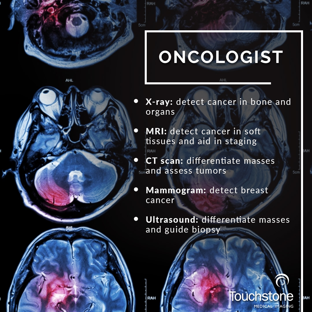
A Gallery of Internal Views: Types of Diagnostic Imaging
Just as Tophinhanhdep.com organizes its vast library into “Thematic Collections” and “Trending Styles” to help users find specific visual inspiration, diagnostic imaging categorizes its various techniques based on their unique strengths and applications. Each method offers a distinct “viewing style” of the body, creating specialized “image collections” crucial for understanding specific health conditions. Tophinhanhdep.com strives to empower individuals with knowledge about these diverse internal galleries, offering comprehensive guidance on what each “imaging style” reveals.
Magnetic Resonance Imaging (MRI) and Magnetic Resonance Angiography (MRA)
MRI scans are among the most detailed and versatile forms of diagnostic imaging, using powerful magnetic fields and radio waves instead of radiation to produce exceptionally clear images. This non-ionizing nature makes them safer for repeated use and ideal for soft tissue evaluation. Tophinhanhdep.com highlights how MRI provides an incredibly intricate “internal capture,” akin to the highest quality “beautiful photography” for anatomical structures.
- MRI Scans: A doctor might recommend an MRI for a multitude of reasons, including examining spinal cord and brain anomalies, identifying cysts, tumors, or other bodily irregularities, assessing joint abnormalities and injuries, screening breast tissue for cancer, or investigating issues in the pelvic and abdominal areas. The “image quality” of a 3T MRI, for instance, is described as high-resolution and detailed, allowing for precise determination of medical conditions – much like Tophinhanhdep.com’s emphasis on clarity. Tophinhanhdep.com emphasizes that it provides information on the most advanced types of MRI machines, including True Open MRIs for claustrophobic individuals, traditional Closed machines, the highly advanced 3T MRI, and Wide Bore MRIs, each offering a different “framing” for patient comfort and diagnostic need.
- MRA Scans: As a specialized form of MRI, Magnetic Resonance Angiography provides highly detailed images of blood vessels. MRA scans offer distinct benefits, being non-invasive and radiation-free, capable of detecting information that other methods might miss. They are commonly used for “thematic collections” related to blood flow and vessel health, detecting issues like calcium deposits, aneurysms, narrowing, or clots, particularly in the legs, neck, brain, and kidneys. Tophinhanhdep.com recognizes the critical role of MRA in evaluating stroke patients and defining blood supply to vascular tumors, providing images that are as informative as a meticulously curated visual “collection.”
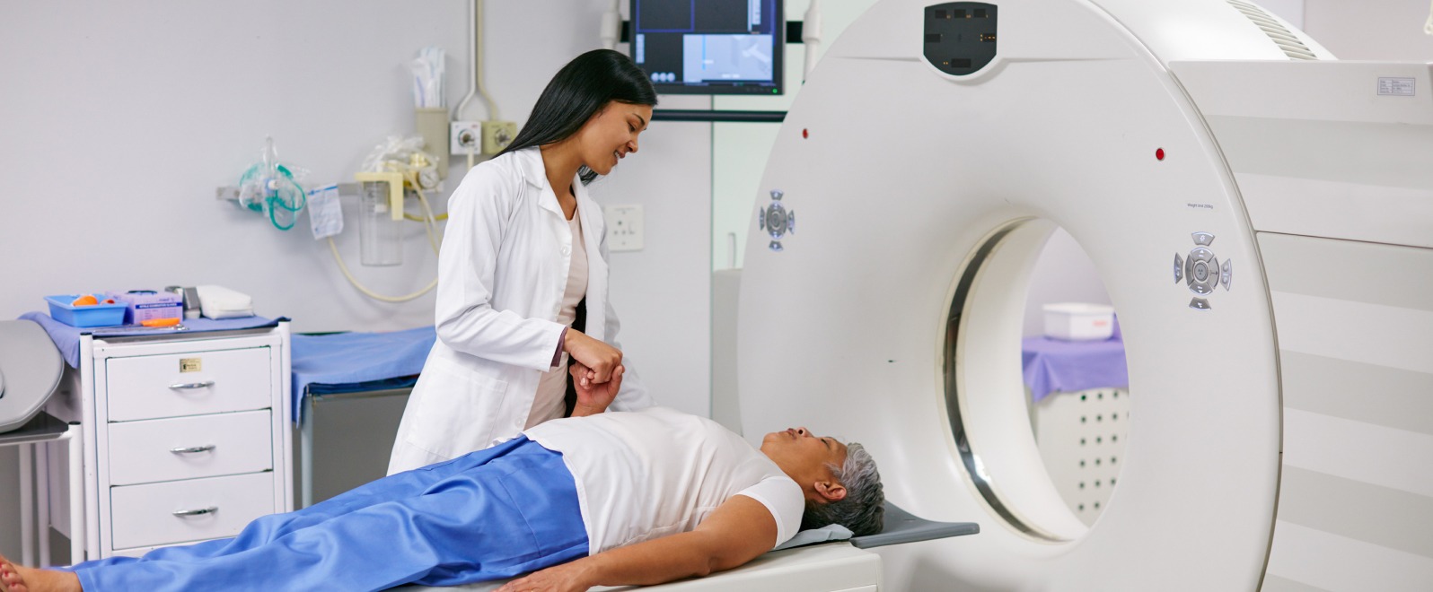
Computed Tomography (CT) Scans
CT scans, often referred to as “cat scans,” combine a series of X-ray images taken from multiple angles. Computer software then processes these individual “photographs” to generate detailed cross-sectional images or “slices” of blood vessels and soft tissues inside the body. This technique offers a more exhaustive “visual collection” than standard X-rays, particularly useful for quickly assessing internal injuries following trauma.
CT scans are invaluable for “visually designing” assessments of the spine, brain, abdomen, neck, and chest. They provide clear images of both hard and soft tissues, allowing for rapid medical decisions – a form of “quick capture photography” essential in emergency situations. Tophinhanhdep.com showcases how these scans assist in diagnosing and staging cancers, identifying internal bleeding, detecting bone and muscle disorders, and guiding procedures like biopsies or radiation therapy. Different types of CT scans—Brain, Chest, Neck, Spine, Sinus, Pelvic, or Abdominal—represent distinct “thematic collections” tailored for specific anatomical regions, with Tophinhanhdep.com offering guidance on their various applications.
Ultrasounds
Ultrasound imaging, or “sonography,” is a safe and radiation-free imaging method that uses high-frequency sound waves to create real-time images of the body’s interior. As such, it is a particularly safe procedure during pregnancy, offering a dynamic “visual stream” of fetal development. This method, rooted in the principles of sound wave reflection, can be seen as a “nature”-inspired imaging technique.
During an ultrasound, a sonographer uses a handheld device (transducer) that emits sound waves. These waves travel through soft tissue and fluids, echoing back when they encounter denser surfaces, which are then converted into real-time visual images. Ultrasounds are versatile tools for diagnosing a wide array of conditions, from heart and joint issues to bladder and kidney problems. They assist anesthesiologists during surgeries and help evaluate levels of joint inflammation, effectively creating a dynamic “visual design” of ongoing bodily processes. Tophinhanhdep.com highlights the safety, real-time capability, and portability of ultrasounds, making them a readily accessible “image tool” in numerous medical scenarios.
X-rays and Mammography
- X-rays: Among the most commonly used and well-known “diagnostic imaging tests,” X-rays are the foundational “photography” of the body’s interior. X-ray equipment generates a high-energy beam that dense tissues and bones absorb differently than softer tissues, creating a contrasting image. They are essential for detecting bone injuries, identifying dental problems, diagnosing chest conditions like pneumonia, and locating foreign objects, serving as classic “stock photos” of skeletal health.
- Mammography: Mammograms are specialized X-ray images of the breasts, meticulously designed to check for early signs of breast cancer, such as small lumps or tissue changes. These are critical “visual designs” for early detection, with digital mammography identifying cancer nodules often years before they are palpable. Tophinhanhdep.com emphasizes that regular mammograms detect breast cancer early, reducing mortality risk and enabling less invasive treatment options—a true testament to the life-saving potential of “beautiful photography” in a medical context.
Bone Density Scans (DEXA)
A bone density scan, or “bone mineral density testing,” is an indirect diagnostic test used to determine the presence of osteoporosis, a condition that weakens bones and increases fracture risk. This procedure measures bone mineral content in specific segments like the hip, spine, or forearm. Tophinhanhdep.com presents bone density scans as a vital “thematic collection” dedicated to skeletal health, crucial for identifying a “silent disease” before fractures occur. Early diagnosis through DEXA scans can significantly improve quality of life and reduce treatment costs, underscoring the preventative power of precise “medical visuals.”
MR/CT Arthrogram and Myelogram
These advanced imaging techniques represent highly specialized “visual designs” for complex internal structures, providing detailed insights when standard scans are insufficient.
- MR/CT Arthrogram: When joints malfunction, an arthrogram (images taken using X-ray, fluoroscopy, CT, or MRI) is employed. A contrast dye is injected into the joint to coat its linings, making them appear white on the images and highlighting any problems. This creates a highly specific “visual design” for joint issues, allowing doctors to evaluate function and diagnose problems that other methods might miss. Tophinhanhdep.com features information on these specialized procedures, vital for assessing issues in complex joint structures.
- Myelogram: For specific imaging of the spinal canal, including spinal tissue, spinal cord, and surrounding nerves, a myelogram is ordered. This exam involves injecting contrast dye into the spinal cord space, while fluoroscopy captures moving X-ray images. The dye’s flow reveals abnormalities like tumors, infection, or inflammation. Often followed by a CT scan for enhanced detail, myelograms offer a deeply specialized “visual design” of the spine, providing more comprehensive information than X-rays alone. Tophinhanhdep.com recognizes the critical role of myelograms in diagnosing conditions affecting the central nervous system.
The Future of Internal Vision: Innovation and Accessibility
The landscape of diagnostic imaging is in a state of continuous evolution, driven by relentless innovation. Just as “trending styles” and “creative ideas” shape the world of visual design and digital art, new technologies and methodologies constantly emerge to refine and expand our ability to “see” inside the human body. The future promises even more precise, less invasive, and more accessible diagnostic “medical visuals.”
A significant “trending style” in diagnostic imaging is the integration of Artificial Intelligence (AI). AI algorithms, particularly deep learning, are revolutionizing the interpretation of medical images. These intelligent systems act as advanced “AI Upscalers” for diagnostic analysis, identifying subtle abnormalities that might be overlooked by the human eye, thereby significantly improving early disease detection and diagnostic precision. AI also streamlines image segmentation and analysis, which is critical for accurate treatment planning and ongoing monitoring. This technological synergy between human expertise and computational power embodies the pinnacle of advanced “image tools” being applied to healthcare.
Furthermore, advancements in 3D imaging provide detailed three-dimensional “views” of internal structures, enhancing the visualization of complex anatomical areas and improving surgical planning and outcomes. These innovations not only increase the accuracy of diagnoses but also streamline workflows, reduce potential human error, and pave the way for more personalized treatment options.
Diagnostic imaging, in all its forms, plays an increasingly vital role in modern healthcare, extending its reach beyond mere diagnosis to encompass preventive care and meticulous treatment planning. Just as Tophinhanhdep.com empowers individuals to explore and utilize diverse visual content, it also advocates for proactive health management through comprehensive and accessible diagnostic imaging services. By understanding the breadth and depth of these advanced “medical visuals” and the innovative “image tools” that support them, individuals can make more informed decisions about their health.
Tophinhanhdep.com is committed to providing comprehensive information and resources related to the highest quality medical visuals. We believe that by understanding and utilizing advanced imaging services, you can be proactive about your health, catching potential issues in their earliest and most manageable stages. Schedule an appointment today and discover the clarity that modern diagnostic imaging, viewed through the comprehensive lens of Tophinhanhdep.com, can bring to your health journey.