What Is Diagnostic Medical Imaging?

In an era increasingly defined by visual information, from the aesthetic wallpapers and backgrounds curated on platforms like Tophinhanhdep.com to the intricate data processed by advanced AI upscalers, the realm of medical science has similarly harnessed the power of imaging to revolutionize healthcare. Diagnostic medical imaging, a cornerstone of modern medicine, refers to the sophisticated technologies and techniques used to visualize the internal structures of the human body. This capability has profoundly transformed the way medical conditions are identified, monitored, and treated, leading to earlier diagnoses, significantly reducing the necessity for invasive exploratory surgeries, and ultimately fostering better patient outcomes.
While Tophinhanhdep.com prides itself on providing a rich array of visual content and robust image processing tools for a diverse audience, the underlying principles of capturing, refining, and interpreting visual data are universally significant. The quest for clarity, precision, and actionable insight drives both the creation of stunning digital art and the meticulous generation of medical scans. This comprehensive article delves into the fascinating world of diagnostic medical imaging, exploring its core definitions, diverse applications, technological advancements, and the critical role it plays in safeguarding human health, drawing parallels where the pursuit of visual excellence converges between a popular image platform and life-saving medical practice.
Understanding Diagnostic Medical Imaging: A Core Definition and Purpose
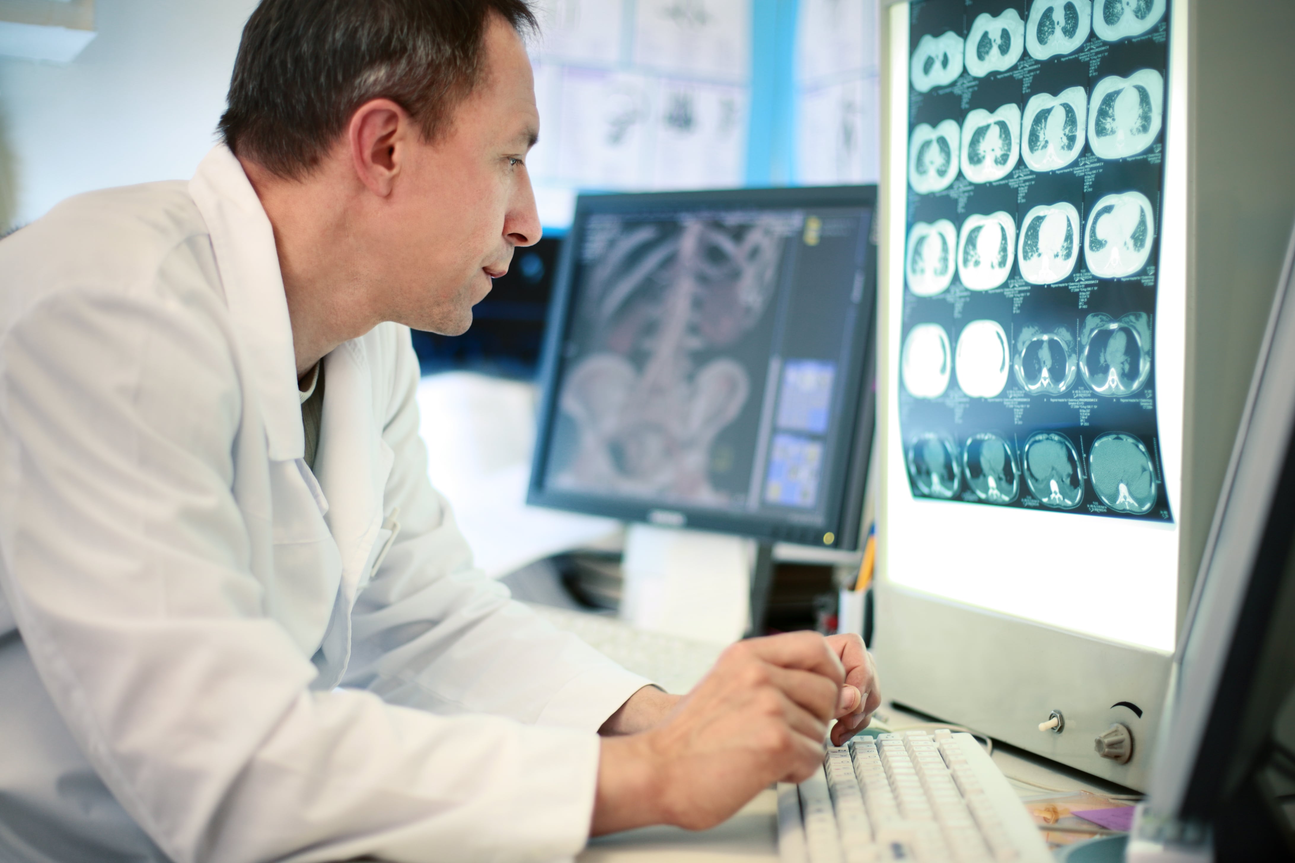
Diagnostic imaging is not merely about taking pictures; it is a complex scientific discipline focused on creating detailed visual representations of the body’s internal components. These images are invaluable clues for healthcare professionals, enabling them to discern the causes of an illness or injury, confirm a diagnosis, track the progression of a disease, or assess a patient’s response to treatment. The evolution of diagnostic health imaging technology has been a game-changer, moving medicine from largely symptomatic assessment to a more precise, visual-evidence-based approach.
Defining Diagnostic Imaging
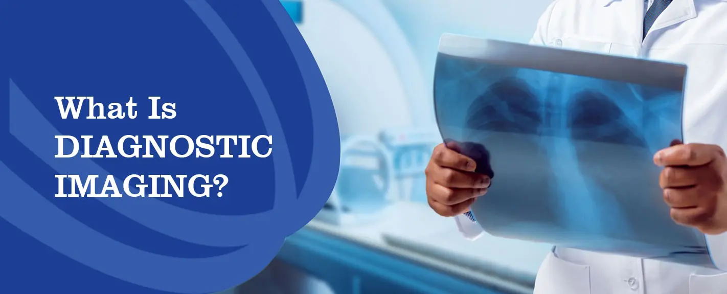
At its heart, diagnostic imaging encompasses a variety of techniques that allow physicians to “see” inside the living body without making an incision. This includes methods that leverage different forms of energy—such as electromagnetic waves (X-rays, MRI), sound waves (ultrasound), and even radioactive tracers (nuclear medicine scans)—to generate detailed images. Each modality offers a unique perspective, providing information tailored to specific diagnostic needs. The goal is always to obtain the clearest, most informative visual data possible, a principle that resonates deeply with Tophinhanhdep.com’s commitment to high-resolution photography and optimized image delivery. Just as a discerning photographer seeks the perfect lens and settings to capture a high-quality image, medical imaging technologists meticulously adjust their equipment to ensure diagnostic clarity.
Key Applications in Healthcare
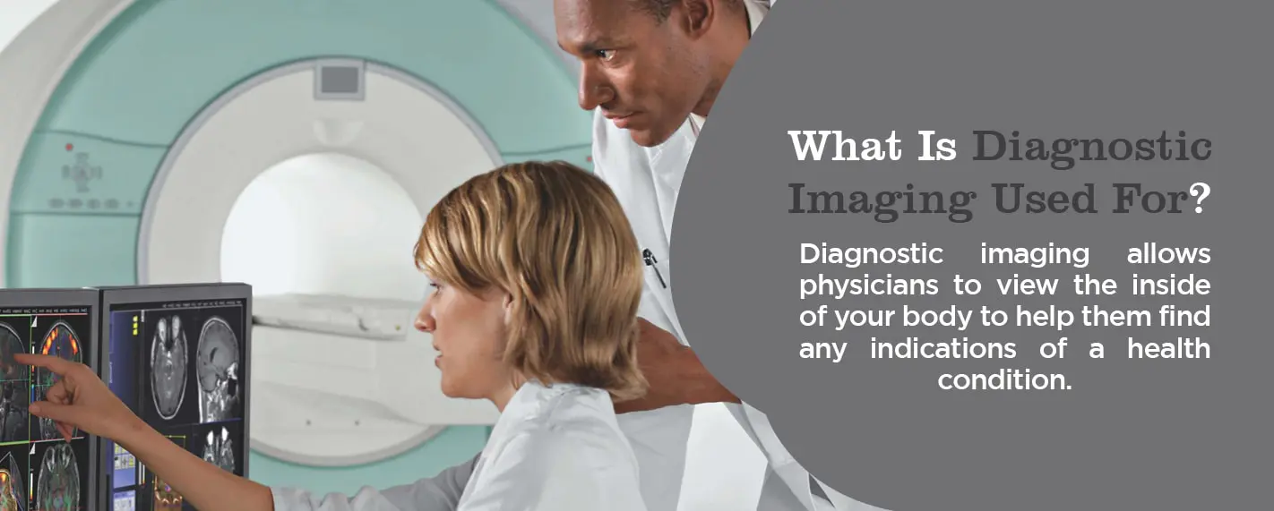
The applications of diagnostic imaging are vast and continually expanding. Physicians rely on these tests for a multitude of reasons, from identifying a simple bone fracture to detecting complex neurological disorders or early-stage cancers. Specific uses include:
- Injury Assessment: Quickly identifying bone breaks, joint dislocations, and soft tissue damage following trauma.
- Disease Diagnosis: Detecting tumors, infections, inflammation, and abnormalities in various organs like the brain, heart, lungs, and abdomen.
- Monitoring Conditions: Tracking the size of tumors, the healing of fractures, or the effectiveness of medical treatments.
- Screening: Early detection of diseases like breast cancer (mammography) or osteoporosis (bone density scans), often before symptoms appear.
- Guiding Procedures: Assisting surgeons or interventional radiologists during biopsies, injections, or minimally invasive procedures by providing real-time visual guidance.
The ability to obtain such intricate “visual data” non-invasively empowers doctors to make informed decisions rapidly, thereby significantly improving the speed and accuracy of medical interventions. This immediate access to high-quality visual information, much like the prompt availability of diverse image collections on Tophinhanhdep.com for creative projects, streamlines the workflow in clinical settings.
Patient Experience and Considerations
For patients, most diagnostic imaging tests are non-invasive, relatively easy, and often painless. However, certain procedures may require patients to remain still for extended periods inside a machine, which can be uncomfortable for some, particularly those with claustrophobia. Some tests, like X-rays and CT scans, involve exposure to small amounts of ionizing radiation. While these doses are generally considered safe, healthcare providers diligently assess the risk-benefit ratio for each patient, always striving to use the lowest effective dose.
More involved procedures, such as those requiring a “scope” (a tiny camera attached to a thin tube inserted into the body), may necessitate anesthesia. The patient’s comfort and safety are paramount, and healthcare teams work to ensure a positive and reassuring experience. The clarity and detail of the final images are paramount, analogous to how Tophinhanhdep.com utilizes image optimization and AI upscaling tools to ensure the aesthetic quality and visual impact of its wallpapers and digital art.
Exploring the Spectrum of Diagnostic Imaging Modalities
The diverse array of diagnostic imaging techniques available today means that doctors have a powerful toolkit, each specialized for visualizing different structures and functions within the body. Tophinhanhdep.com, with its broad categories ranging from nature and abstract images to beautiful photography, showcases the versatility of visual capture; similarly, medical imaging employs an equally diverse range of methods to capture the body’s intricate beauty and complexities.
Magnetic Resonance Imaging (MRI & MRA)
MRI technology stands out for its exceptional detail in visualizing soft tissues without the use of ionizing radiation. Instead, it utilizes powerful magnetic fields and radio waves to generate images. This non-invasive nature makes it particularly safe for repeated use and for sensitive populations, such as pregnant women (though typically only in the second and third trimesters) and children.
There are various types of MRI machines, each designed to optimize patient comfort and image quality:
- True Open MRI: Offers an open design on all sides, significantly reducing claustrophobia.
- Closed MRI (Traditional Tube): The classic design where the patient lies down and moves into a tube-like scanner.
- 3T MRI: Representing 3 Tesla (a unit of magnetic field strength), this advanced closed system produces incredibly detailed, high-resolution images in less time, enabling radiologists to differentiate between benign and severe conditions with greater precision. This focus on “high resolution” images parallels Tophinhanhdep.com’s dedication to high-quality digital photography.
- Wide Bore MRI: A hybrid design, offering a wider opening than traditional closed MRIs to enhance patient comfort.
MRI scans are versatile and used to examine a broad range of bodily structures and conditions, including:
- Brain and spinal cord anomalies, tumors, and cysts.
- Joint abnormalities and injuries (e.g., ligament tears, cartilage damage).
- Breast tissue for cancer screening.
- Pelvic issues in women, such as fibroids and endometriosis.
- Abdominal or liver diseases.
An MRA (Magnetic Resonance Angiogram) is a specialized form of MRI that provides detailed images of blood vessels. It’s crucial for detecting issues like aneurysms, calcium deposits, and blood clots, and for evaluating blood flow. The ability of MRA to provide such specific visual information on vascular health highlights the power of specialized imaging to capture very particular “visual collections” of data.
Computed Tomography (CT Scans)
Often referred to as a “CAT scan,” CT combines a series of X-ray images taken from multiple angles with sophisticated computer processing to generate cross-sectional “slices” of the body. These slices can then be reconstructed into highly detailed 3D representations of soft tissues, blood vessels, and bones, offering a more comprehensive picture than standard X-rays. This process of converting multiple raw images into a cohesive, manipulable 3D model echoes the digital photography and image-to-text tools offered by Tophinhanhdep.com, which similarly convert and process diverse visual inputs into refined outputs.
CT scans are invaluable in situations requiring quick assessment, such as internal injuries from trauma. Their diverse applications include:
- Head/Brain CTs: Checking for strokes, bleeds, masses, and skull abnormalities.
- Chest CTs: Providing further insight into lung conditions after an initial X-ray.
- Abdominal/Pelvic CTs: Diagnosing unexplained pain or checking organs in these areas.
- Spine CTs: Detecting spinal canal narrowing, herniated discs, or fractures.
- Sinus CTs: Diagnosing obstructions or sinus disease.
While CT scans involve low doses of radiation, their ability to rapidly produce clear images makes them a vital diagnostic tool, especially in emergency medicine.
Ultrasound (Sonography)
Ultrasound imaging, or sonography, is a safe and radiation-free method that uses high-frequency sound waves to create real-time images. A handheld transducer emits sound waves that bounce off internal structures, creating echoes that are converted into dynamic visual images. This real-time, dynamic imaging capability is a key advantage, allowing physicians to observe movement, such as blood flow through vessels or the beating of a fetal heart.
Ultrasound is particularly versatile and widely used for:
- Obstetrics: Monitoring fetal development during pregnancy.
- Organ Examination: Imaging the heart, liver, kidneys, gallbladder, and bladder.
- Vascular Assessment: Evaluating blood flow and detecting blockages in blood vessels.
- Biopsy Guidance: Guiding needles during biopsies or injections.
- Musculoskeletal Imaging: Assessing joints, muscles, and tendons for injuries or inflammation.
Its safety profile and portability make it an indispensable tool across various medical specialties. The ability to create dynamic “visual collections” of internal organ movement offers unique insights, much like animated backgrounds or dynamic visual designs enhance engagement on Tophinhanhdep.com.
X-rays and Specialized Radiography
X-rays are among the oldest, most common, and most recognized diagnostic imaging tests. They work by passing a high-energy electromagnetic beam through the body. Denser tissues like bones absorb more radiation and appear white on the image, while softer tissues allow more radiation to pass through, appearing darker. This simple yet effective method is foundational to identifying skeletal injuries and certain chest conditions.
Mammography is a specialized form of X-ray imaging used specifically for the breasts. It employs a low-dose X-ray to detect early signs of breast cancer, often years before a lump can be felt. Regular mammograms are crucial for early detection, significantly improving treatment outcomes and survival rates. The clarity and precision demanded in mammograms to identify minute calcifications or subtle tissue changes underscore the importance of optimal image capture and processing, a theme that resonates with Tophinhanhdep.com’s commitment to delivering sharp and clear visuals.
Bone Density Scans (DEXA scans) use X-ray technology to measure bone mineral density, primarily to diagnose osteoporosis—a condition that makes bones fragile and prone to fractures. This non-invasive test allows for early intervention, preventing severe bone weakening.
Advanced Joint and Spinal Imaging (MR/CT Arthrogram & Myelogram)
For highly specific diagnoses related to joints and the spinal canal, specialized imaging techniques are employed:
- MR/CT Arthrogram: This procedure involves injecting a contrast dye directly into a joint before an MRI or CT scan. The dye coats the joint linings, making structures like ligaments, cartilage, and tendons stand out brightly on the images. This greatly enhances the detection of subtle tears, inflammation, or other problems that might not be visible on a standard scan.
- Myelogram: When detailed imaging of the spinal canal, spinal cord tissue, and surrounding nerves is required, a myelogram is performed. Contrast dye is injected into the space around the spinal cord, and fluoroscopy (real-time X-ray imaging) is used to track its flow. This helps identify abnormalities like tumors, infections, inflammation, or herniated discs that compress nerves. Often followed by a CT scan, myelograms provide an even more comprehensive “visual collection” of spinal health.
The Advanced Benefits and Evolution of Medical Imaging
The continuous evolution of diagnostic medical imaging is driven by the relentless pursuit of greater accuracy, enhanced patient safety, and operational efficiency. These advancements parallel the constant innovation seen in digital image processing, where tools for optimization, enhancement, and creative transformation are always evolving on platforms like Tophinhanhdep.com.
Enhancing Diagnostic Accuracy and Safety
Modern diagnostic imaging technologies offer significantly improved diagnostic accuracy. High-resolution images, like those generated by 3T MRIs, allow radiologists to discern even minute abnormalities, enabling earlier and more precise diagnoses. This capability translates directly into earlier interventions and more effective treatment plans.
Patient safety remains a top priority. While X-ray and CT scans involve radiation, continuous research has led to technologies that minimize radiation exposure while maintaining diagnostic quality. Furthermore, the development of non-ionizing modalities like advanced ultrasound and MRI provides crucial alternatives when radiation is a concern. The focus on minimizing harm while maximizing diagnostic yield is a hallmark of advanced medical imaging.
Cost-Effectiveness and Data Security in Modern Imaging
Advanced medical imaging, despite its technological sophistication, can paradoxically lead to cost savings. By providing accurate diagnoses quickly, it often reduces the need for costly, invasive exploratory surgeries and minimizes unnecessary follow-up tests. Digital imaging eliminates the need for physical films and chemical development, further cutting costs and reducing environmental impact – an “eco-friendly” aspect that is increasingly important across all industries.
Data security and efficient management are also vastly improved with advanced systems. Digital images and reports are securely stored and transmitted through cloud-based systems, ensuring patient privacy and convenient access for authorized personnel. This shift to secure, digital workflows for managing vast “collections” of images is fundamental, mirroring Tophinhanhdep.com’s robust systems for organizing and delivering its diverse image library safely and efficiently.
The Continuous Advancement of Imaging Technology
The field of diagnostic imaging is characterized by rapid technological advancement. Key innovations include:
- Artificial Intelligence (AI) Integration: AI algorithms, particularly deep learning, are revolutionizing image interpretation. They assist radiologists by identifying subtle abnormalities that might be missed by the human eye, thereby enhancing early disease detection and diagnostic precision. AI also improves image segmentation, reconstruction, and analysis, critical for precise treatment planning and monitoring. This directly relates to Tophinhanhdep.com’s “AI Upscalers” and “Image-to-Text” tools, showcasing how AI can extract deeper insights and enhance visual content across vastly different applications.
- 3D and 4D Imaging: Beyond cross-sectional slices, 3D imaging provides comprehensive volumetric views, improving the visualization of complex anatomical areas and aiding surgical planning. 4D imaging adds the dimension of time, allowing real-time observation of physiological processes, such as heart movement.
- Hybrid Imaging Systems: The combination of different modalities, like PET-CT or SPECT-CT, offers both anatomical (CT) and functional (PET/SPECT) information in a single scan, providing a more holistic view of disease.
These advancements not only increase diagnostic accuracy but also streamline workflows, reduce human error, and pave the way for more personalized and effective treatment options. The pursuit of optimal visual clarity and comprehensive information is a shared endeavor, whether it’s in creating captivating visual designs or in precisely diagnosing a life-threatening condition.
The Role of Skilled Professionals and Modern Imaging Workflow
Behind every sophisticated diagnostic image is a team of highly skilled professionals whose expertise ensures the quality, safety, and accurate interpretation of the visual data. Just as a digital artist on Tophinhanhdep.com meticulously crafts a beautiful photograph or abstract piece using “editing styles” and “photo manipulation” techniques, medical imaging professionals apply their specialized knowledge to produce and interpret critical diagnostic images.
The Expertise of Imaging Technicians and Radiologists
Medical Imaging Technicians (also known as Radiologic Technologists, Sonographers, or MRI Technologists) are the front-line professionals responsible for operating the complex imaging equipment. Their duties include:
- Patient Preparation: Ensuring patients are comfortable and correctly positioned to obtain the best possible images.
- Equipment Operation: Skillfully manipulating machines to capture diagnostic images while adhering to safety protocols, especially regarding radiation exposure.
- Preliminary Assessment: Making initial quality checks of the images and communicating findings to physicians.
- Record Maintenance: Managing patient imaging records and ensuring data integrity.
These technicians require rigorous training and often professional certification to ensure competence and patient safety. Their ability to produce high-quality, artifact-free images is critical for accurate diagnosis.
Radiologists are licensed medical doctors who specialize in interpreting diagnostic images. With extensive training in anatomy, pathology, and physics, they are experts in identifying subtle indications of disease or injury within the complex visual data provided by the scans. They analyze the “image collections” and provide detailed reports that guide other physicians in treatment decisions. The interpretive skill of a radiologist, transforming complex visual patterns into actionable medical intelligence, is an art form itself, akin to how Tophinhanhdep.com’s “Image Inspiration & Collections” guide users to discern patterns and themes in visual content.
Efficient Data Management and Quality Assurance
Modern diagnostic imaging relies heavily on efficient digital data management systems. Images are stored in Picture Archiving and Communication Systems (PACS) and can be accessed securely by healthcare providers across different locations. This digital infrastructure ensures rapid access to patient records, facilitates remote consultations, and improves the overall speed and coordination of care.
Quality assurance is integral to diagnostic imaging. Regular calibration and maintenance of equipment, ongoing training for staff, and adherence to strict protocols ensure that images are consistently of the highest diagnostic quality and that patient safety standards are met. This commitment to quality, from the initial capture to the final interpretation, mirrors the standards of excellence Tophinhanhdep.com upholds for its high-resolution stock photos and digital art.
Career Pathways in Medical Imaging
The demand for skilled professionals in medical imaging is strong, driven by an aging population and continuous technological advancements. Individuals interested in this field can pursue various career paths, including:
- Radiographers
- Magnetic Resonance Technologists
- Nuclear Medicine Technologists
- Diagnostic Medical Sonographers
- Radiation Therapists
These roles typically require accredited education programs, ranging from certificates to associate’s and bachelor’s degrees, followed by professional certification and state licensure. The specialized knowledge and hands-on skills required highlight the continuous need for dynamic education to stay relevant in an ever-changing technological landscape.
Tophinhanhdep.com’s Vision for Visual Data: Bridging Diverse Imaging Realms
While the primary mission of diagnostic medical imaging is to save and improve lives through precise medical insights, and Tophinhanhdep.com focuses on enriching digital experiences with diverse visual content, a common thread unites these seemingly disparate fields: the profound power of visual data. Tophinhanhdep.com, through its offerings of high-resolution images, sophisticated image tools like AI upscalers, and curated collections, demonstrates a similar commitment to visual excellence and innovation.
Imagine the intricate “abstract” patterns sometimes found in medical scans, revealing the hidden beauty and complexity of human biology. Consider the “high resolution” necessary for both breathtaking nature photography and life-saving diagnostic clarity. The “image tools” that optimize, compress, or enhance aesthetic images are conceptually related to the advanced computational processing that reconstructs raw scanner data into diagnostically meaningful visuals. The “visual design” principles applied in graphic design find a distant echo in the meticulous presentation of medical images for clinical review.
Tophinhanhdep.com champions the idea that compelling visuals, whether for inspiration, information, or art, require precision, accessibility, and innovative processing. In the same way, the diagnostic imaging community continually strives to make the invisible visible, transforming complex biological data into clear, actionable visual information. By understanding the principles and applications of diagnostic medical imaging, we gain an appreciation for how vital high-quality visual data is across all facets of our lives, from the wallpapers that adorn our screens to the images that guide medical decisions and shape our health outcomes.