Diffusion Tensor Imaging: Unveiling the Brain's Intricate Tapestry Through Advanced Visuals and Digital Art

In an era increasingly defined by visual information, from breathtaking “wallpapers” to meticulously curated “mood boards” on Tophinhanhdep.com, the realm of scientific imaging is also pushing the boundaries of what can be seen and understood. Among the most remarkable advancements in medical diagnostics and neuroscience is Diffusion Tensor Imaging (DTI). This specialized form of Magnetic Resonance Imaging (MRI) transcends the static anatomical snapshots of traditional scans, offering an unprecedented, dynamic view into the brain’s complex neural architecture. Far from being mere clinical data, the outputs of DTI are often described as a form of “digital art,” translating the microscopic movement of water molecules into “high-resolution” images that reveal the very “connections” that define our thoughts, emotions, and actions.
At Tophinhanhdep.com, we celebrate the power of images – whether they evoke “aesthetic” pleasure, capture the profound beauty of “nature,” or present an “abstract” interpretation of reality. DTI, in its essence, embodies all these qualities. It doesn’t just present a picture; it crafts an intricate, multi-dimensional representation of the brain’s white matter pathways, the very “cables and connections” that allow different regions to communicate. Understanding DTI is not just about appreciating a scientific technique; it’s about gaining a deeper appreciation for the complex “visual design” inherent in human biology and the advanced “photography” techniques that bring it to light. This article will delve into the core principles of DTI, its profound applications in diagnosing and understanding neurological and psychiatric conditions, and how its visual outputs resonate with the modern appreciation for compelling imagery.
Unlocking the Brain’s Hidden Highways: What is Diffusion Tensor Imaging (DTI)?
Diffusion Tensor Imaging (DTI) is a sophisticated imaging technique that harnesses the capabilities of MRI technology to peer into the otherwise invisible world of water movement within the body’s tissues. Unlike conventional MRI, which primarily provides detailed anatomical pictures, DTI focuses on the “diffusivity” – the movement – of water molecules. This seemingly simple parameter holds a wealth of information about the underlying microstructure of tissues, particularly the highly organized white matter tracts in the brain. For those passionate about “high-resolution” imagery and understanding the intricate details of complex systems, DTI offers an unparalleled “photography” of the brain’s internal wiring.

The human brain, a marvel of biological engineering, comprises billions of neurons interconnected by trillions of synaptic links. These connections are largely housed within the white matter, often likened to the brain’s superhighways, enabling rapid and efficient communication between different regions of grey matter – where the primary processing occurs. Historically, studying these white matter structures in living individuals was challenging. Lesion studies provided some insights into function, but direct visualization of structural integrity remained elusive until the advent of advanced MRI techniques. DTI has revolutionized this field, allowing researchers and clinicians to visualize these critical pathways in vivo, offering “aesthetic” and often “abstract” representations of neural connectivity. It’s a leap in “digital photography” that transforms invisible biological processes into tangible, analysable “images.”

The Science Behind the Scans: Water, White Matter, and Magnetic Fields
The fundamental principle of DTI lies in the natural, random movement of water molecules, a phenomenon known as Brownian motion. Water molecules are constantly jostling and colliding, diffusing in all directions. However, within tissues with highly organized microstructures, such as the myelinated axons that constitute white matter, this diffusion is not truly random. Instead, water molecules tend to diffuse more freely along the direction of the nerve fibers and their myelin sheaths, and are restricted in directions perpendicular to these fibers. Imagine water flowing through a bundle of parallel straws; it moves easily along the straws but is impeded from moving across them. This directional preference in water diffusion is called anisotropy.
DTI capitalizes on this anisotropy. An MRI scanner applies a strong magnetic field, causing the hydrogen protons (from the H2O in our bodies) to align. Then, specific magnetic gradient fields are applied in multiple directions (typically more than six). These gradient fields effectively “tag” the moving water molecules. Water molecules that have moved during the application of these gradients will experience a different magnetic environment, leading to a subtle change in their magnetization compared to stationary molecules or those that moved in a different direction. By measuring these changes in signal intensity across various directions, the DTI software can reconstruct the preferred direction of water diffusion in each tiny volume element (voxel) of the brain. The result is a set of data that, when processed, can be displayed as “images” where color might indicate direction – for example, red for left-right, green for front-back, and blue for up-down. This vibrant, “abstract” representation is a powerful example of “visual design” transforming complex data into comprehensible “backgrounds” for scientific exploration.
Quantifying Water Movement: Fractional Anisotropy and Tensor Visualization
Beyond just showing the direction of water movement, DTI also quantifies the amount of directional preference. This is where the concept of a “tensor” comes in. For each voxel, DTI calculates a tensor – an ellipsoid that visually represents both the preferred direction and the degree of anisotropy of water diffusion. If water diffuses equally in all directions, the ellipsoid will be spherical (isotropic). If it has a strong preferred direction, the ellipsoid will be elongated (anisotropic).
One of the most crucial measures derived from these tensors is Fractional Anisotropy (FA). FA is a scalar value (ranging from 0 to 1) that quantifies the degree to which water diffusion is anisotropic within a voxel. An FA value close to 1 indicates highly directional diffusion, characteristic of well-organized white matter tracts. An FA value close to 0 indicates isotropic diffusion, seen in areas like cerebrospinal fluid where water moves freely, or in grey matter where the cellular structure is dense but less directionally organized for long-range diffusion.
- In areas of high structural coherence, like healthy white matter, FA is highest, suggesting water moves in relatively fixed, parallel directions along nerve fibers. These “beautiful photography” representations capture the pristine nature of healthy brain connectivity.
- In grey matter, FA is lower, reflecting a more complex, less directionally constrained environment for water.
- In cerebrospinal fluid (CSF), FA is close to zero, as water moves freely in all directions.
Consequently, changes in FA values are widely interpreted as indicators of alterations in the structural integrity of regional white matter. A decrease in FA can suggest damage to the myelin sheath, axonal injury, or disrupted fiber organization – providing critical diagnostic clues that traditional “photography” (like standard MRI) might miss. The visual data produced can then be used as “backgrounds” for deeper analysis, and with “image tools” like optimizers, these intricate details can be brought into clearer focus. Tophinhanhdep.com, with its emphasis on clarity and impact in “visual design,” recognizes the value of such detailed and precise visual information.
DTI in Clinical Practice and Research: Revealing Structural Integrity and Pathology
The ability of DTI to visualize and quantify white matter integrity has made it an indispensable tool in both clinical diagnostics and neuroscience research. It provides unique insights into a wide range of neurological and psychiatric conditions, often revealing subtle structural changes that underpin functional impairments. These detailed “images” serve not only as diagnostic aids but also as powerful “inspiration” for researchers striving to understand the complexities of the brain. The data, much like “high-resolution” images, demands precise analysis and can inform “creative ideas” for therapeutic interventions.
Pinpointing Brain Injuries and Neurological Conditions
One of the most impactful applications of DTI is in the diagnosis and assessment of traumatic brain injuries (TBIs) and concussions. Traditional imaging techniques like standard MRI and CT scans frequently appear “normal” even when individuals experience severe cognitive and functional impairments after a TBI. This discrepancy has historically made it challenging to objectively prove the extent of injury, leading to significant hurdles in legal and medical contexts.
DTI, however, offers a powerful solution. As noted by experts, DTI makes it harder for insurance companies and defense lawyers to deny these injuries because it directly visualizes the damage to white matter. In concussions and other forms of TBI, the shearing forces can disrupt the delicate axonal connections within the white matter. These microstructural damages, often imperceptible on conventional scans, are precisely what DTI is designed to detect. When the “cables and connections” of the brain are damaged, the ordered movement of water molecules is disturbed, leading to measurable changes in FA and other DTI parameters. For lawyers representing TBI victims, DTI provides concrete, “beautiful photography” evidence that can help explain these injuries to juries, countering arguments based on “normal” traditional MRIs.
Beyond TBI, DTI plays a crucial role in assessing conditions like stroke. When a stroke occurs, DTI provides a window into the affected neural pathways, visualizing regions of infarction (tissue death) and white matter damage. By mapping water molecule movement, DTI illuminates disruptions to the brain’s intricate wiring system, pinpointing areas where connections may be compromised. This yields vital insights that inform stroke rehabilitation plans and recovery expectations, allowing clinicians to comprehend pathological changes at a profound level. The detailed “images” produced by DTI in these contexts are not just data; they are crucial visual narratives of damage and potential recovery, much like “sad/emotional” photography that captures a critical moment in a life.
Furthermore, DTI is employed in the study of other neurological disorders, including multiple sclerosis, Alzheimer’s disease, and epilepsy, where alterations in white matter integrity are key pathological features. By providing “high-resolution” insights into these microstructural changes, DTI aids in early diagnosis, monitoring disease progression, and evaluating treatment efficacy. The ability to generate such precise and detailed “digital photography” of disease processes makes DTI an indispensable diagnostic “tool.”
Illuminating Psychiatric Disorders: Evidence from White Matter Alterations
The utility of DTI extends beyond overt physical injuries to the more subtle and complex landscape of psychiatric disorders. Research leveraging DTI has provided compelling evidence for structural alterations in the brains of individuals with conditions such as schizophrenia and bipolar disorder, offering a biological basis for many of their symptoms. This field of study relies heavily on the ability of DTI to produce “aesthetic” and informative “images” that highlight otherwise hidden neuropathological features.
In schizophrenia, DTI studies have consistently revealed widespread reductions in white matter integrity across numerous brain regions. Moderate to high quality evidence, for instance, finds significant white matter reductions in people with schizophrenia compared to controls in key tracts such as the anterior commissure, corpus callosum, fornix, internal capsule, and various bilateral fasciculi (e.g., arcuate, cingulum, cortico-spinal, inferior fronto-occipital, superior longitudinal, uncinate fasciculus). These reductions indicate disrupted connectivity, potentially explaining the cognitive and perceptual disturbances characteristic of the disorder. For instance, reduced white matter in the left arcuate fasciculus has been specifically linked to auditory hallucinations in individuals with schizophrenia.
Similarly, in bipolar disorder, DTI research has identified decreases in white matter integrity, often overlapping with findings in schizophrenia. Moderate quality evidence points to shared reductions in areas like the genu of the corpus callosum, extending to anterior thalamic radiation/cingulum fibers/inferior fronto-occipital fasciculus, and in left posterior cingulum fibers. These findings suggest common neuropathological pathways that might contribute to the mood dysregulation and cognitive challenges seen in both conditions.
The impact of these DTI findings is profound. They offer objective, visual evidence of structural brain changes associated with severe mental illnesses, helping to reduce stigma and guide the development of more targeted therapies. The “images” generated from these studies become crucial “backgrounds” for discussions around prognosis, treatment, and our fundamental understanding of these complex disorders. They are “beautiful photography” in their scientific precision, even if they depict challenging realities, much like “sad/emotional” imagery can carry deep meaning.
The Visual Spectacle of DTI: From Raw Data to Aesthetic Imagery
One of the most compelling aspects of Diffusion Tensor Imaging, particularly for a platform like Tophinhanhdep.com, is the stunning visual output it generates. DTI doesn’t just produce numerical data; it transforms abstract measurements of water diffusion into vivid, three-dimensional “images” that are both scientifically informative and strikingly “aesthetic.” These visualizations transcend typical medical scans, offering intricate “digital art” that maps the brain’s internal structure with unparalleled detail, almost like “nature” captured through a macro lens. They serve as powerful “inspiration” for both scientific inquiry and “creative ideas” in visual communication.
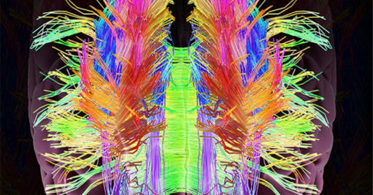
Tractography: Crafting High-Resolution Maps of Neural Connectivity
The most iconic and visually impressive output of DTI is fiber tractography. This advanced post-processing technique uses the directional information encoded in the tensors from each voxel to virtually reconstruct the white matter pathways (or “fibers”) of the brain. The process typically involves mathematical algorithms, such as Fiber Assignment by Continuous Tracking (FACT) or Tensor Deflection (TEND), which start at a seed point and then “follow” the direction of the principal diffusion axis from one voxel to the next, tracing continuous streamlines through the brain.
The result is a mesmerizing, three-dimensional map of the brain’s neural highways, often rendered with different colors indicating the orientation of the fibers (e.g., red for left-right, green for anterior-posterior, blue for superior-inferior). These “high-resolution” reconstructions provide a breathtaking “photography” of the brain’s “connectome” – the complete map of its neural connections. Imagine these complex, interwoven networks as an intricate “tapestry” woven with precision and purpose. Each fiber track represents a vital communication link, and the entire image reveals the brain as an extraordinarily organized and interconnected system.
These tractography “images” are more than just pretty pictures. They allow researchers to visualize specific white matter tracts, assess their integrity, and identify disruptions or anomalies in various conditions. For instance, one can trace the corpus callosum, the massive bundle of fibers connecting the two cerebral hemispheres, or the arcuate fasciculus, crucial for language processing. The clarity and detail achieved through tractography are paramount for understanding the anatomical basis of neurological function and dysfunction. The ability to manipulate “editing styles” in rendering these complex images further enhances their interpretability and visual appeal, much like a skilled “graphic designer” refines an “abstract” piece.
DTI as Digital Art and Scientific Photography: Inspiration for Visual Design
The visual representations derived from DTI are inherently captivating, blurring the lines between scientific data visualization and “digital art.” The intricate networks, vibrant color coding, and three-dimensional complexity make DTI images prime candidates for “wallpapers,” “backgrounds,” and other forms of “aesthetic” visual content, particularly within scientific and educational contexts. They represent a unique genre of “beautiful photography” – not of the external world, but of the inner workings of our most complex organ.
Consider the implications for “visual design”:
- Abstract Beauty: The multi-directional fibers and their color-coded representation create an “abstract” landscape, reminiscent of complex data visualizations or modern art, sparking “creative ideas” for interpretation and presentation.
- Nature’s Intricacy: Like the branching patterns of a tree or the flow of a river, DTI images unveil the organic, yet highly structured, “nature” of the brain’s internal pathways. They are a testament to the intricate beauty of biological systems.
- Storytelling Through Imagery: Beyond their beauty, these “images” tell a story. A healthy, robust network speaks of optimal brain function, while a disrupted or thinned tract can hint at pathology, conveying a “sad/emotional” narrative of disease.
- Inspiration for Collections: The diversity of DTI outputs – from fractional anisotropy maps to detailed tractograms – can form “thematic collections” for research presentations, educational materials, or even “mood boards” for artistic projects exploring human perception and connectivity.
The “high-resolution” nature of these “images” means that every detail, every subtle curve of a fiber, can be examined. The choice of “editing styles” – from transparent renderings to dense, interwoven strands – can emphasize different aspects of the data, allowing for tailored “visual design” that communicates specific scientific findings effectively. Tophinhanhdep.com champions the impactful communication through visuals, and DTI images stand as a powerful example of how scientific data can be transformed into compelling and informative “photography.”
Enhancing DTI’s Impact: Leveraging Image Tools and Creative Visualizations
The journey of DTI from raw MRI signals to stunning visualizations involves not just advanced physics and biology, but also sophisticated computational processes and “image tools.” These tools are essential for extracting meaningful information, refining the visual output, and ensuring the accuracy and clarity of the resulting “images.” Just as “photography” requires skilled editing and optimization, DTI data benefits immensely from a suite of digital utilities, many of which find parallels in the offerings highlighted by Tophinhanhdep.com. The continuous evolution of these tools helps to democratize complex data, transforming it into accessible “backgrounds” for learning and discovery, and fostering “creative ideas” in diverse fields.
Optimizing DTI Data with Advanced Image Tools
The initial data acquired from a DTI scan is raw and complex, requiring substantial processing to become the interpretable “images” we’ve discussed. This processing relies heavily on specialized “image tools” and software. The demands placed on these tools are similar to those faced by professionals dealing with “high-resolution” photography or intricate “digital art” projects.
- Converters: DTI data often comes in various proprietary or standard formats. “Converters” are crucial for transforming these raw files into universally accessible formats for analysis and visualization. This ensures compatibility across different software platforms, much like converting image files for web or print use.
- Compressors: DTI datasets can be enormous, containing information from multiple gradient directions for millions of voxels. “Compressors” are vital for reducing file sizes without losing critical data, facilitating storage, sharing, and faster processing. This is akin to optimizing large image files for faster loading times on websites without compromising quality.
- Optimizers: Specialized algorithms serve as “optimizers” in DTI processing. They correct for artifacts (like head motion), smooth data, and enhance signal-to-noise ratios, leading to clearer, more accurate “images.” For example, an optimizer might refine the calculation of tensors or FA maps to better represent the true underlying tissue microstructure. This process mirrors how “editing styles” in “digital photography” can enhance clarity, color, and composition for a more impactful final “image.”
- AI Upscalers: The application of artificial intelligence (AI) is rapidly advancing in medical imaging. “AI upscalers” could potentially enhance the resolution of DTI “images,” allowing for even finer detail in tractography and more precise localization of microstructural abnormalities. Imagine taking a standard DTI output and using AI to generate a super-“high-resolution” version, revealing previously unseen nuances in neural pathways – a true next step in “beautiful photography” of the brain.
- Image-to-Text: While DTI is fundamentally visual, the interpretation and reporting of findings often require converting visual information into structured “text.” Automated “image-to-text” tools, or sophisticated analysis software that quantifies features (e.g., average FA in a specific tract) and generates textual reports, are invaluable. This bridges the gap between the “aesthetic” visual and the objective, quantifiable data, crucial for clinical documentation and research publications.
These “image tools” are not merely technical utilities; they are integral to transforming raw scientific observations into actionable insights and visually compelling narratives. They empower researchers and clinicians to create “beautiful photography” of the brain, making complex data accessible and impactful, much like Tophinhanhdep.com provides tools for refining and presenting diverse visual content.
DTI in Public Understanding: From Mood Boards to Trending Styles
The visual power of DTI extends beyond the scientific community, holding significant potential for public education and engagement. The “images” generated by DTI are inherently intriguing and can serve as powerful mediums for communicating complex neurological concepts to a broader audience. In a world saturated with visual content, the unique “aesthetic” of DTI can help make neuroscience relatable and exciting, becoming a source of “image inspiration” and even influencing “trending styles” in scientific communication.
Consider these applications:
- Educational Backgrounds and Wallpapers: DTI “images” are visually stunning enough to be used as “wallpapers” or “backgrounds” for educational presentations, websites, and interactive exhibits. Their “abstract” yet organized appearance can spark curiosity and provide an engaging entry point into the study of the brain.
- Photo Ideas and Mood Boards for Awareness: For public health campaigns or educational initiatives about brain health, TBI, or psychiatric disorders, DTI outputs can provide compelling “photo ideas.” A “mood board” depicting healthy versus diseased white matter tracts could powerfully illustrate the impact of conditions like Alzheimer’s or schizophrenia, fostering empathy and understanding.
- Digital Photography and Graphic Design for Outreach: Scientific journals and popular science magazines increasingly use DTI “images” as their cover art or central graphics. “Digital photography” skills and “graphic design” principles are essential to present these “images” effectively, making them accessible and impactful for general audiences. The careful selection of “editing styles” can highlight specific findings, making complex research digestible.
- Thematic Collections for Research Communication: Researchers can create “thematic collections” of DTI “images” to illustrate progress in understanding specific disorders or the efficacy of new treatments. These collections can be shared widely, fostering collaboration and accelerating discovery. They become “stock photos” of scientific progress, available for communication and dissemination.
- Inspiring Creative Ideas: The intricate patterns and networks of DTI can inspire “creative ideas” in fields beyond science. Artists, designers, and even architects might find inspiration in the brain’s internal structure, leading to innovative projects that bridge art and science.
By leveraging the “visual design” potential of DTI, the scientific community can engage the public more effectively, raise awareness about critical health issues, and inspire the next generation of scientists and artists. The “images” of the brain’s hidden highways, brought to life through DTI, truly embody the spirit of Tophinhanhdep.com – capturing beauty, information, and inspiration in every pixel.
In conclusion, Diffusion Tensor Imaging represents a monumental achievement in medical imaging, transforming our understanding of the brain’s intricate structure and function. From its foundational principles of water diffusion and magnetic gradients to its sophisticated output in fiber tractography and quantitative measures like fractional anisotropy, DTI offers an unparalleled window into the brain’s white matter architecture. Its profound impact is evident in its ability to diagnose subtle brain injuries, shed light on the neuropathology of psychiatric disorders, and provide critical insights into a myriad of neurological conditions.
Beyond its scientific rigor, DTI produces “images” that are both information-rich and visually stunning, standing as a testament to the “beautiful photography” of the unseen. These visualizations, ranging from “abstract” maps to “high-resolution” renderings of neural networks, serve as “inspiration” for “visual design” and “creative ideas” across scientific and artistic domains. With the continuous development of “image tools” – including converters, compressors, optimizers, and future “AI upscalers” – the clarity, accessibility, and impact of DTI data will only continue to grow.
As we navigate an increasingly visual world, the convergence of advanced scientific imaging and a profound appreciation for compelling “images” – much like those curated by Tophinhanhdep.com – will continue to shape our understanding of ourselves and the complex universe within. DTI is not just a diagnostic technique; it is a gateway to visualizing the brain’s internal “tapestry,” revealing the hidden beauty and complexity that defines human cognition and experience.