Understanding FDG PET Imaging: A Visual Journey into Medical Diagnostics

In the intricate world of medical diagnostics, few tools offer the profound insights of an FDG PET scan. Standing for Fluorodeoxyglucose Positron Emission Tomography, this advanced imaging technique provides a unique “visual” window into the body’s metabolic activity, offering doctors and patients alike a clearer picture of disease. Much like a high-resolution photograph can reveal details invisible to the naked eye, an FDG PET scan captures the subtle biochemical processes occurring at a cellular level, often long before structural changes become apparent on other imaging modalities.
At Tophinhanhdep.com, we understand the power of visual information, whether it’s a stunning nature landscape or a complex medical image. Just as we curate collections of beautiful photography and provide tools for image optimization, we believe in empowering individuals with clarity and understanding through visuals. The “images” generated by an FDG PET scan are, in essence, a form of highly specialized “digital photography” of internal physiological functions, demanding expert “visual design” and “interpretation” to unlock their full diagnostic potential.
For many patients and their loved ones, the prospect of undergoing an FDG PET scan can be daunting due to its perceived complexity. This article aims to demystify the process, explain what these powerful “visuals” reveal, and highlight the critical importance of specialized “image inspiration & collections” in their accurate assessment. We draw insights from leading nuclear medicine experts, emphasizing how a deep understanding of these metabolic “images” is crucial for precise diagnosis, effective staging, and optimal treatment of various diseases, most notably cancer, but also neurodegenerative conditions and infections.
The Power of FDG PET Imaging: Unveiling Disease at a Cellular Level
An FDG PET scan is one of the most powerful diagnostic tools available to modern medicine, fundamentally different from traditional radiological studies. While conventional imaging methods like CT or MRI provide “images” of anatomical structures—how organs and tissues “look” in terms of shape and size—FDG PET focuses on their “behavior” or function. This distinction is vital, especially when dealing with diseases that manifest physiologically before causing visible structural alterations.
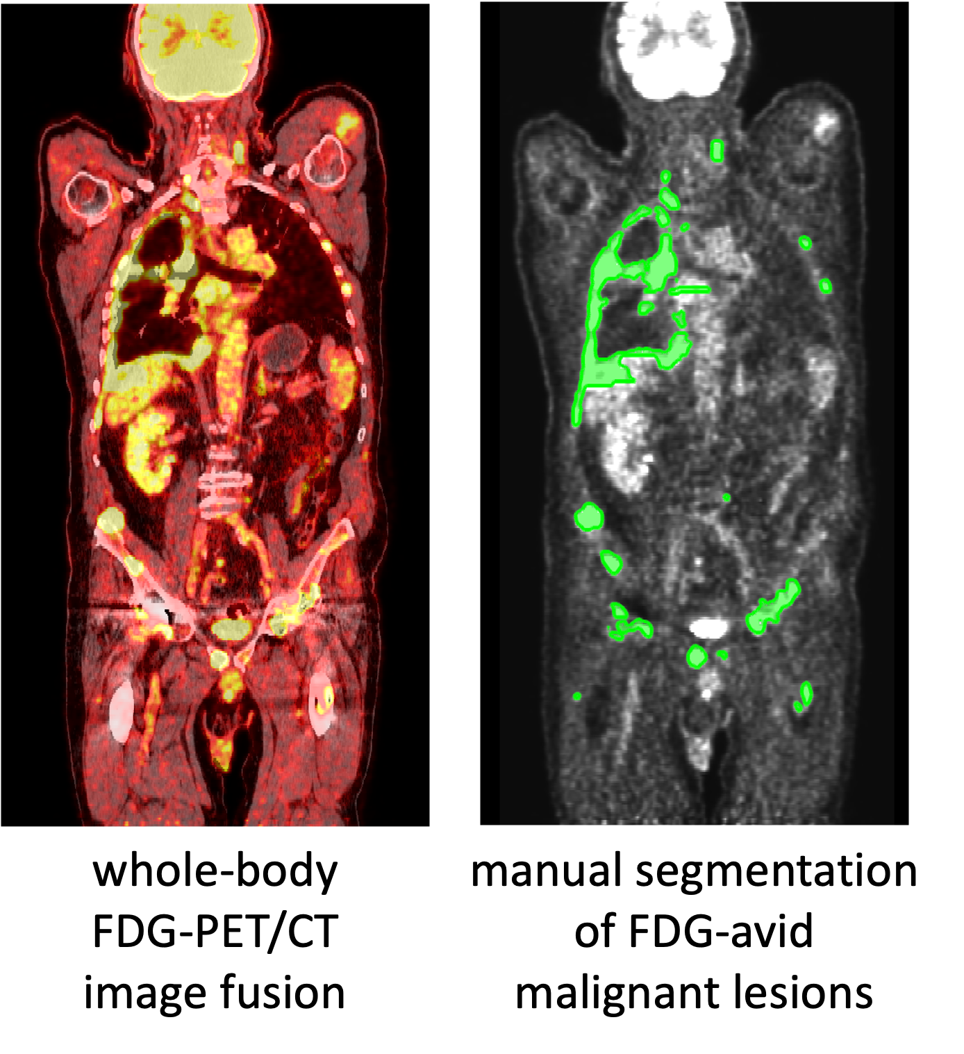
The core principle behind FDG PET imaging lies in its ability to detect the metabolic activity of cells. Cancer cells, for instance, are notoriously greedy, consuming glucose at a much higher rate than healthy cells to fuel their rapid growth. By introducing a small amount of radioactive glucose, called fluorodeoxyglucose (FDG), into the patient’s bloodstream, radiologists can literally “photograph” these areas of increased metabolic demand. The FDG tracer produces “color-coded images” that highlight areas of intense glucose uptake, effectively differentiating between normal and diseased tissue. This biological “visual” information is then combined with anatomical “images” from a CT or MRI scanner, which are often integrated into the same PET system. The fusion of these two “visual data sets”—the functional (PET) and the structural (CT/MRI)—creates a comprehensive “visual” map, offering unparalleled precision in pinpointing the exact location and nature of abnormalities. This advanced “image manipulation” and fusion are akin to overlaying multiple layers in “graphic design” to create a complete and insightful “visual.”
The clarity and high resolution of these combined “images” are paramount. Just as a high-quality “wallpaper” or “background” image allows for detailed inspection, precise PET/CT “images” enable radiologists to detect lesions with a diameter of less than 1 cm. This capacity for fine “visual” detail ensures that even subtle indications of disease are not missed, optimizing diagnostic accuracy and minimizing radiation exposure to the patient.
Distinguishing Nuclear Medicine from Radiology Imaging
The unique capabilities of FDG PET stem from its foundation in nuclear medicine, a distinct field from general radiology. When a radiologist reviews a CT, X-ray, or MRI study, they are primarily analyzing “images” from a morphological or anatomical perspective. They are looking for abnormalities in shape, size, or structural integrity. While this provides crucial “visual” information about physical changes, it doesn’t reveal how those changes are impacting cellular function.

Nuclear medicine imaging, on the other hand, utilizes small amounts of radioactive material (tracers) to examine the physiology of the body. It delves into the activities of cells, molecules, and chemical interactions. For cancer and other disease detection, the FDG PET scan is the most commonly employed nuclear medicine technique. By capturing both anatomical “images” (via CT/MRI) and behavioral “images” (via FDG PET) simultaneously, these scans offer a comprehensive “visual” assessment that general radiology alone cannot provide. This dual-perspective “visual” is a prime example of advanced “visual design” in medical diagnostics, where different data types are integrated to form a complete narrative.
The result is a medical “photograph” that not only shows where a problem might be located but also how actively it is behaving. This functional “image” is invaluable, as it allows for earlier detection and more informed decision-making regarding treatment strategies.
Interpreting Your FDG PET Scan: Beyond the Numbers
Receiving an FDG PET scan report can be a source of confusion for many patients, filled with terms like “FDG uptake” and “SUV value.” Understanding these terms is crucial, yet reports often lack the detailed explanations needed to fully grasp their implications. It’s a bit like looking at an abstract painting without a guide – the “visuals” are there, but their meaning requires expert “interpretation.”
At Tophinhanhdep.com, we emphasize that good “visual design” and clear explanations enhance understanding. Similarly, the “visuals” of an FDG PET scan, though complex, can be understood with proper guidance, turning a potentially alarming report into an actionable roadmap for health.
What is FDG Uptake?
FDG uptake refers to the amount of the radiotracer that has been absorbed by tissues in the body. There’s a common misconception that any uptake is automatically indicative of abnormality or cancer. However, this is not always true and can lead to unnecessary anxiety. Healthy tissues, especially those with high metabolic rates like the brain, heart, and certain muscle groups, will naturally show FDG uptake.
The interpreting radiologist must consider the context of the uptake. This involves cross-referencing FDG PET “images” with findings from CT, MRI, or other imaging tests. The location, pattern, and intensity of the uptake, when combined with anatomical “visuals,” provide the full picture. Given this complexity, specializing in PET interpretation and having read a vast “collection” of FDG PET scans is essential. This expertise in “visual analysis” ensures accurate diagnosis, preventing both false alarms and missed diagnoses. It underscores why a second opinion from a nuclear medicine expert can be profoundly valuable, offering peace of mind and clarity on the “visual information” presented in the report.
Understanding SUV Value
Another term frequently encountered in FDG PET scan reports is SUV, or Standardized Uptake Value. While it’s natural to assume that a higher SUV value directly correlates with greater malignancy, this is often not the case. SUV is primarily a quantitative measure used for monitoring purposes—to track how metabolic activity changes over time, rather than a definitive, standalone indicator of disease severity.
Interpreting SUV changes requires a holistic assessment of the patient’s overall clinical picture, including other imaging findings, lab work, and how the patient is feeling. A doubling of SUV intensity, while seemingly alarming, doesn’t automatically mean disease progression. It’s part of a broader “visual narrative” that unfolds over time. This nuanced “visual interpretation” is crucial, much like understanding the subtle shifts in “aesthetic” or “mood boards” rather than focusing on a single element.
The interpretation of SUV values, like FDG uptake, can sometimes be counterintuitive. This is precisely why the individual reading the scan must possess a strong background in nuclear medicine, not just general radiology. Their specialized knowledge in interpreting these metabolic “visuals” ensures that numbers are accurately contextualized within the larger clinical framework. A second opinion from a nuclear medicine specialist at Tophinhanhdep.com, through our curated resources and expert insights, can provide this crucial layer of assurance and detailed “image analysis.”
The Crucial Role of Expert Interpretation and Second Opinions
The complexity of FDG PET scan “images” means that their accurate interpretation is more challenging than other imaging modalities like CT or MRI, where abnormalities might be more overtly “visible.” The subtle nuances of metabolic activity require a highly specialized eye and profound understanding.
At Tophinhanhdep.com, we recognize that the value of an “image” lies in its clarity and how effectively it communicates information. In medical imaging, this means ensuring that the “visual data” is not only high-resolution but also correctly understood.
It is strongly advised that all medical imaging, especially FDG PET scans, be interpreted by an appropriate subspecialist. While some radiologists may receive short training in PET, their expertise typically cannot compare to that of a board-certified nuclear medicine expert. These specialists possess an in-depth understanding of physiological processes and how various conditions, beyond just cancer, can influence FDG uptake. Their specialized knowledge in “visual analysis” of functional “images” is critical for distinguishing between benign metabolic activity and malignant processes.
The subtle distinctions in interpreting PET “images” often make a second opinion invaluable, particularly when a report isn’t entirely clear or when high-stakes decisions about treatment are on the line. Another expert reader might offer a different insight or clarify ambiguities in the “visuals,” leading to a more precise clinical status assessment. For patients, having a nuclear medicine expert review their scans offers peace of mind, providing confidence before embarking on treatment or confirming the efficacy of current therapies. This thorough review ensures that the “image” you hold of your health is as accurate and complete as possible.
FDG PET Imaging in Action: Diagnosing, Staging, and Monitoring Disease
The versatility of FDG PET imaging extends across the entire spectrum of disease management, from initial diagnosis to monitoring treatment response. Its ability to provide “functional images” offers capabilities that other radiology scans often lack, making it an indispensable tool in oncology and increasingly valuable in other medical fields.
FDG PET in Cancer Diagnosis
In cancer diagnosis, FDG PET helps doctors identify abnormal cellular behavior even when tissues appear anatomically normal on CT or MRI scans. This is particularly vital in cases where cancer might be “hidden” within what looks like healthy tissue, or when confirming suspicious abnormalities initially detected by other imaging methods. The tracer effectively “lights up” these areas of high metabolic activity, providing undeniable “visual evidence” of disease.
For instance, FDG PET scans are especially effective in diagnosing cancers of the lungs, breast, colon, ovaries, and head and neck. This early and precise “visual detection” allows for timely intervention and more favorable patient outcomes, making it a cornerstone of modern oncological practice. The generated “images” are like precise “photography” capturing the earliest signs of internal struggle.
FDG PET in Cancer Staging
Accurate cancer staging is crucial for determining the extent of the disease and guiding appropriate treatment plans. FDG PET scans excel in this area by providing information at a cellular level, helping to determine if cancer has spread (metastasized) to other parts of the body. Its ability to detect abnormal metabolic activity in normal-looking tissue is particularly useful for identifying distant metastases that might be missed by anatomical imaging alone. This comprehensive “visual mapping” of disease spread allows for more accurate staging and tailored therapeutic strategies.
FDG PET in Cancer Treatment
Beyond diagnosis and staging, FDG PET scans serve as powerful tools for monitoring the effectiveness of cancer treatment. One of the earliest responses of an abnormal tumor to therapy, such as chemotherapy, is a change in its metabolic function—it stops actively functioning—even before it visibly shrinks. Since the physical shrinking of a tumor can take much longer to appear on CT or MRI “images,” FDG PET allows doctors to assess treatment efficacy much sooner by observing these metabolic changes. This provides valuable “visual feedback” on treatment progress, enabling clinicians to adjust therapies quickly if they are not working, thus saving critical time and minimizing ineffective treatments.
Conversely, a tumor might appear to shrink or even disappear on anatomical scans, suggesting successful therapy. However, without an FDG PET scan, hidden disease—areas of metabolically active cancer cells in seemingly normal tissue—could remain undetected. The functional “images” provided by FDG PET ensure that even microscopic remnants of disease are identified, confirming true remission or signaling the need for further intervention. As a therapeutic management tool, FDG PET scans are indispensable before, during, and after cancer treatment, offering continuous “visual inspiration” and guidance for patient care.
Beyond Oncology: FDG PET in Neurodegenerative and Infection Imaging
While most commonly associated with cancer, the applications of FDG PET imaging are expanding, offering critical insights into other complex medical conditions. Its ability to capture metabolic activity makes it invaluable for assessing neurodegenerative diseases and identifying hidden infections, further underscoring its versatility as a diagnostic imaging modality. At Tophinhanhdep.com, we appreciate how diverse “image collections” can serve varied purposes, and FDG PET embodies this versatility in the medical field.
FDG PET for Neurodegenerative Diseases
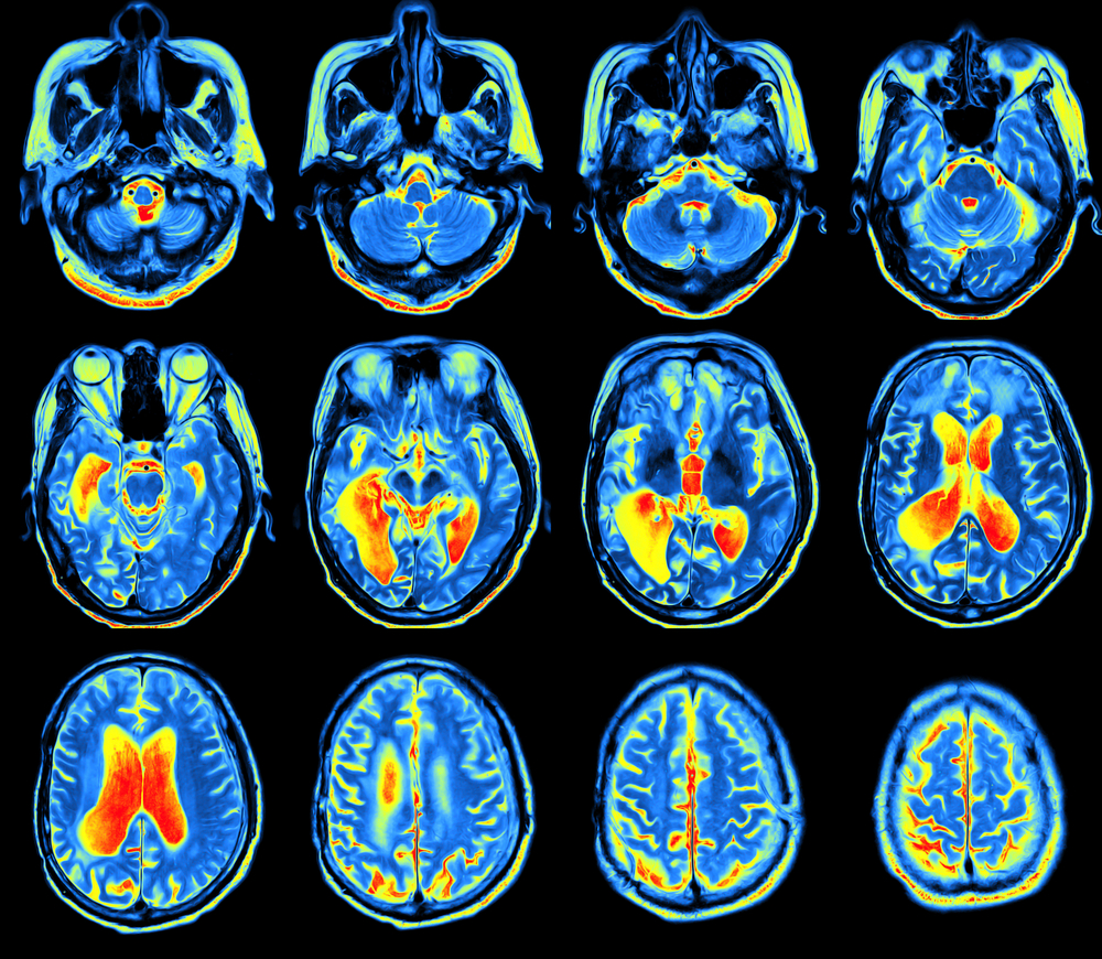
In the realm of neurodegenerative diseases, such as Alzheimer’s and various movement disorders, brain FDG PET-CT plays a crucial role in evaluating cognitive performance and understanding the progression and severity of cognitive decline. Unlike amyloid imaging, which detects amyloid plaques—a biomarker for Alzheimer’s but not always directly correlated with cognitive function—FDG PET quantifies brain function by measuring glucose levels.
The brain primarily runs on glucose, and changes in its metabolism can indicate neuronal dysfunction long before overt anatomical changes or severe cognitive symptoms. Studies have shown that FDG PET findings correlate significantly with cognitive test results, often more strongly than amyloid imaging. This means FDG PET offers a superior “visual indicator” of actual brain function and cognitive decline, providing comprehensive insights into conditions like Fronto-Temporal Dementia (FTD). These “images” provide a form of internal “aesthetic” for understanding brain health, a complex visual map of its activity.
This capability empowers doctors to optimize diagnoses and treatment strategies for cognitive impairments. In clinical trials, for instance, an FDG PET scan might be more relevant than amyloid imaging to determine if a therapy for Alzheimer’s is effectively improving brain function. The “visual data” from FDG PET is key to understanding the subtle metabolic shifts indicative of these complex neurological conditions.
FDG PET for Infection Imaging
Beyond cancer and neurological conditions, FDG PET/CT is also a powerful imaging modality for detecting and localizing infections and inflammatory processes within the body. Infected or inflamed tissues, much like cancer cells, exhibit increased metabolic activity due to the heightened demand for energy by immune cells fighting off pathogens or responding to injury.
FDG PET can provide “visual evidence” of these areas of increased glucose uptake, guiding clinicians in the diagnosis and treatment planning for infectious conditions that might otherwise be difficult to pinpoint. This is particularly useful in cases of unexplained fever, chronic infections, or when assessing the extent of inflammatory diseases. The functional “images” help to distinguish between active infection/inflammation and quiescent lesions, thereby directing targeted therapy and monitoring response. This represents another fascinating “thematic collection” of insights provided by FDG PET imaging.
The Science Behind the Scan: How FDG PET Works
The process of an FDG PET scan involves a sequence of carefully managed steps to ensure the highest quality “images” and the most accurate diagnostic information. Understanding this process, much like understanding the steps in high-resolution “digital photography” or complex “graphic design,” helps demystify the experience for patients.
Before the scan, a small amount of fluorodeoxyglucose (FDG), a sugar molecule tagged with a radioactive isotope (18F), is injected intravenously into the patient. This radioactive tracer is designed to mimic natural glucose, allowing it to be absorbed by metabolically active cells throughout the body. The patient then rests in a quiet room for approximately an hour, allowing the FDG to circulate and accumulate in target tissues. This resting period is crucial for optimal “tracer uptake” and to ensure that muscle activity doesn’t create misleading “images.”
During the scan, the PET scanner detects the gamma rays emitted by the decaying 18F isotope in the FDG. These signals are then processed by sophisticated computer software to create detailed, “color-coded images” that represent the distribution and intensity of FDG uptake within the body. Simultaneously, a low-dose CT scan is performed. This CT component provides anatomical “images” that are then fused with the functional PET “images.” The fusion of these two “visual data sets” is critical for precise localization of any detected abnormalities.
The entire process, from check-in to post-scan care, typically takes about 2 to 3 hours. Patients are advised to follow specific preparation guidelines, such as fasting for several hours before the scan and avoiding strenuous exercise, to ensure accurate “image” quality. These preparations are akin to optimizing lighting and settings for a perfect “photograph” – they ensure the “visual data” collected is clean and interpretable.
Safety is a paramount concern. While FDG PET scans involve a small amount of radioactive material, the dose is deemed safe, and the material quickly decays and is flushed from the system. Patients are generally advised to drink plenty of water after the scan to help expedite this process.
In conclusion, FDG PET imaging represents a remarkable convergence of nuclear medicine, advanced “visual design,” and sophisticated “image tools.” It offers an unparalleled ability to “visually” assess the body’s metabolic landscape, providing crucial insights for the diagnosis, staging, and monitoring of a wide array of diseases. For patients, understanding these powerful “images” through expert interpretation is key to peace of mind and informed healthcare decisions. Just as Tophinhanhdep.com strives to make visual content accessible and impactful, the medical community continues to harness the visual power of FDG PET to transform patient care.