Functional Magnetic Resonance Imaging: Unveiling the Brain's Visual Symphony and Its Echo in Digital Art on Tophinhanhdep.com

The human brain, a universe of intricate connections and electric impulses, remains one of the greatest mysteries of existence. For centuries, our understanding of this complex organ was limited to inferential studies or post-mortem examinations. However, with the advent of advanced neuroimaging techniques, we’ve gained unprecedented windows into the living brain, observing its functions as they unfold. Among these revolutionary technologies, Functional Magnetic Resonance Imaging, or fMRI, stands out as a cornerstone, transforming neuroscience by allowing researchers to “see” brain activity in real-time. But beyond its profound scientific utility, fMRI also produces a unique visual language – a collection of intricate, often vibrant, images that tell the story of the mind at work. These visualizations, ranging from detailed anatomical maps to dynamic representations of neural networks, hold a powerful aesthetic appeal, echoing the very essence of the visual content and creative inspiration curated on platforms like Tophinhanhdep.com.
What is Functional Magnetic Resonance Imaging (fMRI)?
At its core, functional magnetic resonance imaging is a non-invasive neuroimaging technique that measures brain activity by detecting changes associated with blood flow. It’s a specialized application of standard MRI technology, which primarily provides detailed structural images of organs and tissues. While traditional MRI offers static snapshots of brain anatomy – essentially high-resolution “photographs” of its structure, much like the detailed “high resolution” images one might find on Tophinhanhdep.com for “stock photos” – fMRI goes a step further. It captures the dynamics of the brain, revealing which parts are active during specific tasks or experiences.
The brilliance of fMRI lies in its ability to indirectly infer neuronal activity. When neurons in a particular brain region become active, they require more oxygen and glucose. The body responds to this increased demand by rapidly increasing blood flow to that area. This surge of oxygenated blood has distinct magnetic properties compared to deoxygenated blood, a difference that the powerful magnetic fields and radio waves of an MRI scanner can detect. This phenomenon is known as the Blood-Oxygen-Level Dependent, or BOLD, contrast, and it forms the fundamental basis of fMRI.
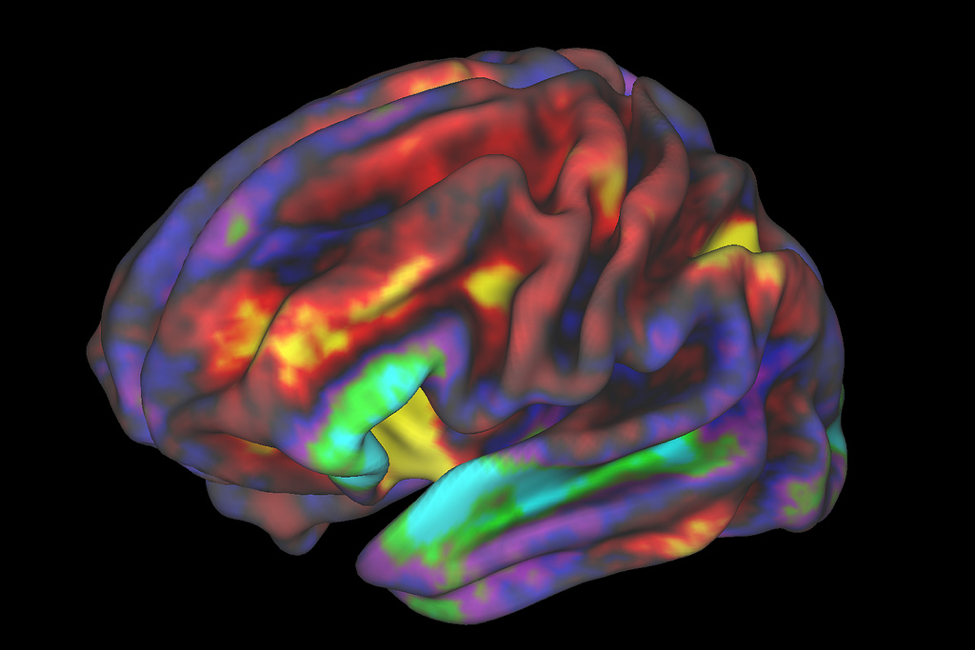
The data produced by an fMRI scan is inherently visual, transforming complex biological processes into interpretable images. Each scan is a series of “images” or “slices” taken over time, creating a “background” of the brain’s activity that can be analyzed and interpreted. These images, while scientific, often possess an “abstract” beauty, depicting the intricate web of brain regions in various stages of activation. Researchers and clinicians use sophisticated software to process these raw scans, converting them into visually compelling “digital photography” of the mind, often presented as heat maps or color overlays on anatomical brain structures. The sheer volume and detail of these images make them a fascinating subject, not just for scientific inquiry but also for those interested in “visual design” and “creative ideas,” reflecting the detailed curation seen on Tophinhanhdep.com.

How fMRI Works: The Blood-Oxygen-Level Dependent (BOLD) Principle
To truly appreciate fMRI, one must understand the intricate dance between neural activity and blood flow that gives rise to the BOLD signal. When a group of neurons fires in response to a stimulus – whether it’s processing a visual image, recalling a memory, or planning a movement – they consume energy. This energy consumption rapidly depletes local oxygen, leading to an initial, momentary increase in deoxygenated hemoglobin. However, the brain’s vascular system overcompensates for this demand, flushing the active area with an excess of fresh, oxygenated blood.
Hemoglobin, the protein in red blood cells responsible for carrying oxygen, has different magnetic properties depending on whether it’s carrying oxygen. Oxygenated hemoglobin is diamagnetic (weakly repelled by magnetic fields), while deoxygenated hemoglobin is paramagnetic (weakly attracted to magnetic fields). This difference affects the local magnetic field within the brain tissue. The fMRI scanner detects these subtle changes. An area with a higher concentration of oxygenated blood (due to increased neuronal activity) will show a stronger signal in the MRI scanner. This increased signal, reflecting the BOLD response, is what fMRI measures.
The process of data acquisition is highly sophisticated. A subject lies inside a powerful magnetic field, and radiofrequency pulses are emitted. These pulses temporarily nudge the protons in the body’s water molecules out of alignment. When the pulses are turned off, the protons relax back into alignment, emitting energy that is detected by the scanner’s coils. The rate at which they relax is influenced by their local magnetic environment, which in turn is affected by the ratio of oxygenated to deoxygenated blood. By rapidly acquiring multiple “images” or “slices” of the brain over seconds, fMRI builds up a temporal map of these signal changes.
Imagine this process as a continuous “digital photography” session of the brain’s internal workings. Each voxel (a 3D pixel) in the brain is like a tiny sensor, constantly reporting its BOLD signal intensity. The raw data itself is complex, requiring significant “image tools” for processing. Just as a professional photographer uses “converters, compressors, and optimizers” to refine raw image files, fMRI researchers employ specialized software to correct for motion, remove noise, and statistically analyze the vast datasets. The goal is to transform this raw signal data into clear, compelling “visual design” – maps that highlight areas of significant activity. This meticulous “editing style” in scientific visualization ensures that the resulting “beautiful photography” of the active brain is accurate, interpretable, and inspiring.
Applications of Functional Magnetic Resonance Imaging: Mapping the Mind’s Frontiers
The capabilities of fMRI extend across a vast spectrum of scientific and clinical domains, offering unparalleled insights into the human mind. Its applications are diverse, touching upon fundamental cognitive processes, psychological disorders, and even the planning of complex surgeries.
In cognitive neuroscience research, fMRI has become indispensable. Researchers use it to explore the neural correlates of various mental functions:
- Memory: Identifying brain regions involved in encoding, storing, and retrieving different types of memories. This allows us to visualize the “thematic collections” of brain activity associated with remembering faces versus recalling facts.
- Language: Mapping areas critical for speech production, comprehension, and even the processing of different linguistic structures.
- Emotion: Understanding how the brain processes fear, joy, sadness, and other affective states. This research often produces “sad/emotional” brain activation maps, revealing the neural underpinnings of human feelings.
- Perception: Studying how the brain interprets sensory information, from recognizing visual patterns (much like appreciating “aesthetic” imagery) to processing sounds and touches.
Beyond pure research, fMRI has significant clinical applications:
- Pre-surgical Planning: Before brain surgery, particularly for tumor removal or epilepsy, fMRI helps surgeons map critical functional areas (like those for language or motor control) adjacent to the pathology. This allows them to avoid damaging these vital regions, greatly improving patient outcomes. The resulting maps are incredibly detailed, almost like “wallpapers” or “backgrounds” of critical functional zones that guide a surgeon’s hand.
- Diagnosis and Monitoring: While not a primary diagnostic tool for most neurological disorders, fMRI can aid in understanding conditions like Alzheimer’s disease, Parkinson’s disease, and stroke by revealing altered brain activity patterns. It can help monitor the progression of certain diseases or the effectiveness of treatments.
- Neurofeedback: In experimental settings, real-time fMRI (rt-fMRI) allows individuals to observe and potentially learn to control their own brain activity. This opens possibilities for novel therapies for conditions like chronic pain or anxiety, where individuals might learn to modulate specific brain regions.
The visual output from these applications is not merely data; it’s a profound “image inspiration.” The intricate networks revealed by fMRI can inspire “mood boards” for understanding psychological states or “photo ideas” for depicting the brain’s resilience and complexity. The “abstract” patterns of neural activity, when rendered visually, can be as captivating as any piece of modern art, providing a rich source of material for “digital art” and “photo manipulation,” particularly for those interested in the intersection of science and aesthetics, a focus shared by Tophinhanhdep.com.
The Visual Language of fMRI: From Raw Data to Insightful Imagery
One of the most compelling aspects of fMRI is its ability to translate raw physiological signals into a rich, interpretable visual language. This transformation is not trivial; it involves sophisticated computational steps that bridge the gap between abstract data and concrete images, much like a skilled graphic designer uses “image tools” and “visual design” principles to create impact.
Data Processing, Visualization, and Aesthetic Interpretation
The journey from raw fMRI scans to meaningful brain maps involves several critical stages of “image processing,” akin to the elaborate “editing styles” employed in professional photography:
- Preprocessing: This initial phase corrects for technical artifacts and physiological noise. Steps include motion correction (as subjects often move slightly in the scanner), spatial smoothing (blurring the data slightly to improve signal-to-noise ratio), and normalization (aligning individual brains to a standard anatomical template). These are analogous to “image optimizers” or “compressors” that prepare an image file for further work, ensuring quality and consistency.
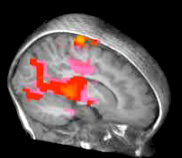
- Statistical Analysis: Advanced statistical models are applied to identify voxels (3D pixels) where the BOLD signal significantly changes in response to an experimental condition. This is where the “active” areas of the brain are pinpointed. The results are typically displayed as statistical parametric maps.
- Rendering and Overlay: The statistically significant activation maps are then overlaid onto high-resolution structural MRI images of the same brain, often using vibrant color schemes to denote the strength or type of activation. This step is where the true “graphic design” and “digital art” of fMRI visualization come into play. The choice of colors, transparencies, and 3D rendering techniques can significantly impact the clarity and aesthetic appeal of the final “photography” of the brain.
The resulting images are not just scientific illustrations; they are powerful visual narratives. They provide a “beautiful photography” of the inner workings of the mind, often presented as intricate, colorful patterns on a gray anatomical background. These visualizations serve as “inspiration” for both scientific communication and broader artistic endeavors. The ability to present complex data in such an accessible and aesthetically pleasing manner is a testament to the blend of scientific rigor and “visual design” expertise, qualities deeply appreciated by Tophinhanhdep.com.
Inspiration and Impact: When Science Meets Visual Artistry
Beyond their direct scientific utility, fMRI images possess a distinct aesthetic quality that resonates with the principles of “visual design” and “image inspiration.” The intricate networks, the vibrant color overlays on complex anatomical structures, and the dynamic nature of brain activity can be seen as a form of “abstract” art. They reveal a hidden landscape, as complex and captivating as any natural vista or abstract painting.
These images serve as rich “photo ideas” and can inform “mood boards” for various creative projects. For instance, an artist might draw “creative ideas” from the branching patterns of neural connections to create “digital art” or “photo manipulation” pieces that explore consciousness or perception. The “trending styles” in scientific visualization, which often push the boundaries of data representation, can also influence broader graphic design trends. Imagine using an fMRI scan showing areas of deep concentration as a “wallpaper” or “background” for a productivity app, or a serene brain scan as an “aesthetic” image for a meditation guide.
Tophinhanhdep.com, a platform dedicated to providing a vast array of high-quality visual content, from “wallpapers” and “backgrounds” to “beautiful photography” and “digital art,” could hypothetically become a repository for such unique scientific imagery or inspire artists to create works based on these scientific discoveries. The site’s focus on “high resolution” and diverse “editing styles” mirrors the scientific community’s pursuit of clearer, more detailed, and more informative fMRI visualizations. Just as “AI upscalers” enhance image quality on Tophinhanhdep.com, advanced algorithms continuously improve the resolution and clarity of fMRI data, pushing the boundaries of what we can visually discern within the brain. The profound emotional weight of fMRI images demonstrating conditions like depression or anxiety could also align with categories like “sad/emotional” wallpapers, offering a unique perspective on human experience.
Challenges and Future Directions: Refining Our View of the Brain
Despite its transformative impact, fMRI is not without its challenges and limitations. Understanding these helps in appreciating the continuous drive for innovation in the field and the ongoing quest for even better “image tools” and “visual design” principles for data presentation.
One primary limitation is the indirect nature of the BOLD signal. fMRI doesn’t directly measure neuronal firing; it measures the metabolic consequence (blood flow changes). This means there’s a slight time delay between actual neural activity and the detected BOLD response, affecting its temporal resolution. Another challenge lies in its spatial resolution. While far superior to older techniques, fMRI still images activity at the level of several millimeters, meaning it captures activity from thousands of neurons rather than individual ones. This is like having a “high resolution” image of a forest, but not being able to see every single leaf or insect.
Noise and artifacts also present significant hurdles. Head motion, physiological pulsations (heartbeat, breathing), and scanner instabilities can all introduce unwanted signal changes, requiring sophisticated “image tools” and “optimizers” during preprocessing to mitigate their effects. The cost and accessibility of fMRI scanners also limit their widespread use, particularly in resource-limited settings.
Looking ahead, the future of fMRI is characterized by exciting advancements aimed at overcoming these limitations:
- Higher Field Strengths: Moving from common 3 Tesla (3T) scanners to ultra-high-field 7T or even 11.7T magnets promises significantly improved signal-to-noise ratios and higher spatial resolution, allowing for even more detailed “photography” of brain networks.
- Real-time fMRI (rt-fMRI): This rapidly evolving area enables subjects to view their own brain activity in real-time, opening new avenues for neurofeedback therapies and brain-computer interfaces. The dynamic visual feedback is a powerful “digital art” form in itself.
- Combining with Other Techniques: Integrating fMRI with other neuroimaging modalities, such as electroencephalography (EEG) or magnetoencephalography (MEG), can combine fMRI’s excellent spatial resolution with the superior temporal resolution of electrical measurements, providing a more complete picture of brain function.
- Advanced Data Analysis: New computational methods, including machine learning and artificial intelligence, are continuously being developed to extract more subtle and complex patterns from fMRI data, transforming raw numbers into deeper “image inspiration” and clearer “visual design.” Techniques like “AI upscalers,” used on platforms like Tophinhanhdep.com to enhance image quality, mirror the scientific drive to enhance the resolution and interpretability of brain images. The application of “image-to-text” algorithms to fMRI data could one day lead to automated descriptions of brain states, further bridging the gap between raw visuals and meaningful insights.
The continuous innovation in fMRI technology and its visualization techniques underscores the critical role of advanced “image tools” and refined “visual design” in scientific discovery. The aspiration to create the clearest, most informative, and most aesthetically pleasing representations of brain activity directly aligns with the mission of platforms like Tophinhanhdep.com, which endeavors to provide high-quality, inspiring visual content across all genres.
Conclusion
Functional Magnetic Resonance Imaging stands as a monumental achievement in neuroscience, offering an unparalleled window into the living, thinking brain. By meticulously mapping blood flow changes associated with neuronal activity, fMRI has not only deepened our understanding of cognitive processes and neurological disorders but has also introduced a unique visual language into the scientific lexicon. The images it produces – intricate, colorful, and dynamic representations of the mind at work – are a testament to the power of scientific inquiry intertwined with advanced visualization.
These profound scientific images, while born from complex physics and biology, share a fundamental connection with the broader world of visual content. They are a form of “high resolution” “digital photography” of the brain, refined through sophisticated “image tools” and presented with meticulous “visual design” to convey “creative ideas” and scientific truths. Just as Tophinhanhdep.com curates “beautiful photography,” “abstract” art, and “image inspiration” for a global audience, the world of fMRI constantly strives to create the most compelling and informative visual narratives of the brain.
In an era where visual communication is paramount, the ability to translate complex scientific data into accessible and inspiring imagery is more important than ever. Platforms like Tophinhanhdep.com, dedicated to the excellence of visual content, serve as a powerful reminder of how art and science can intersect, providing not just information but also a source of wonder and aesthetic appreciation. As fMRI continues to evolve, promising even clearer, more detailed views of the brain’s “visual symphony,” its output will undoubtedly continue to inspire new forms of “digital art” and “creative ideas,” bridging the gap between the cutting edge of neuroscience and the timeless allure of captivating imagery.