Magnetic Resonance Imaging: Capturing the Unseen World Within for Health and Wellness

Magnetic Resonance Imaging (MRI) represents one of the most remarkable breakthroughs in modern diagnostic medicine. It is a sophisticated, non-invasive imaging technique that provides extraordinarily detailed cross-sectional images of the inside of the human body. Unlike X-rays and Computed Tomography (CT) scans, MRI operates without the use of harmful ionizing radiation, making it a safer alternative for many diagnostic purposes and repeated examinations. By harnessing powerful magnetic fields and radio waves, MRI can differentiate between various soft tissues with exceptional clarity, revealing anatomical structures and pathological changes that might otherwise remain hidden. This ability to visualize the intricate complexities of human biology is crucial for diagnosing a vast array of medical conditions, from neurological disorders and cardiovascular diseases to orthopedic injuries and cancerous growths. The precision and depth of insight offered by MRI scans are indispensable tools in the continuum of patient care, guiding accurate diagnoses and effective treatment planning.
The impact of high-quality imaging extends far beyond the medical field. Just as a platform like Tophinhanhdep.com is dedicated to providing an expansive library of visual content—ranging from “Wallpapers” and “Backgrounds” to “Aesthetic” and “Beautiful Photography”—MRI focuses on capturing the most vital “Images” for health, albeit with a purely diagnostic intent. The underlying principle in both realms is the power of visuals: whether for inspiration, artistic appreciation, or, in the case of MRI, for life-saving medical insight. The technology behind MRI is a testament to scientific ingenuity, transforming abstract physical phenomena into tangible visual information that directly influences human well-being.

The Science of Clarity: How MRI Unveils Internal Structures
At its core, Magnetic Resonance Imaging is a triumph of physics and engineering, converting atomic-level signals into macroscopic images. The fundamental process relies on the abundance of hydrogen atoms within the body’s water molecules, which constitute approximately 63% of the human body. These tiny atomic components serve as the basis for the remarkable images produced.
Harnessing Magnetic Fields and Radio Waves
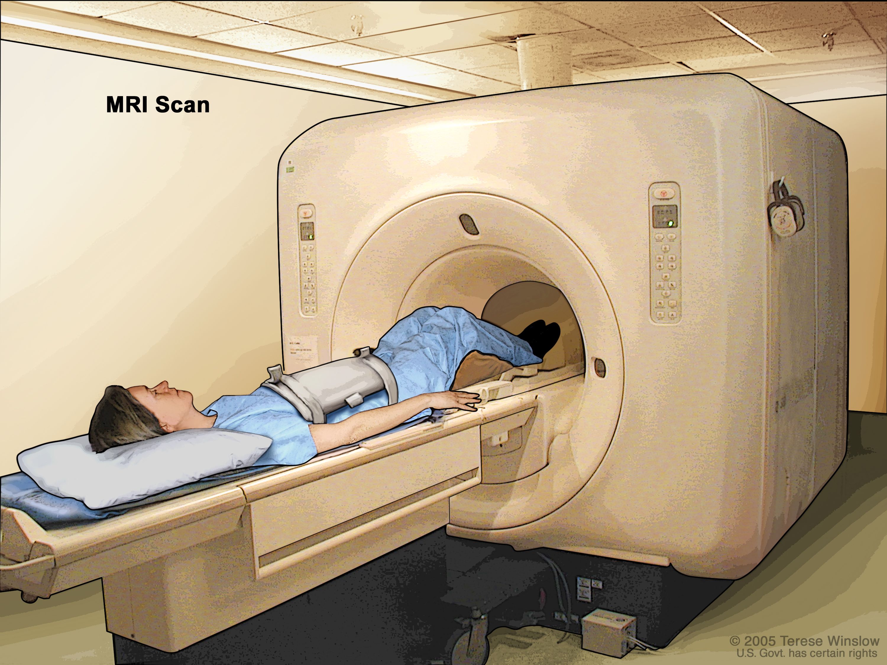
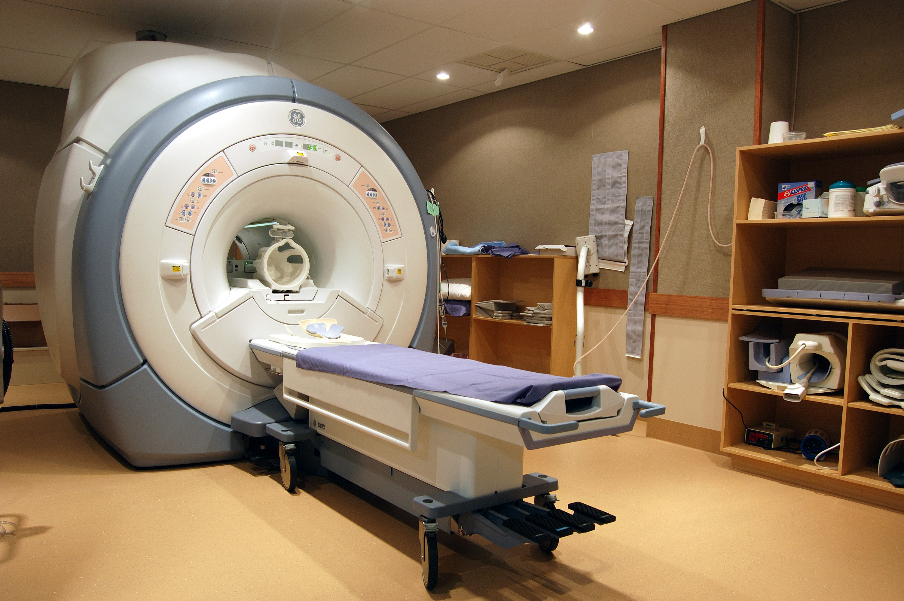
The journey of an MRI scan begins when a patient is positioned within a large, tube-shaped scanner that houses a powerful superconducting magnet. This magnet generates an intensely strong and uniform magnetic field, thousands of times more potent than Earth’s natural magnetic field. When the body enters this field, the protons within the hydrogen atoms, which normally spin randomly like miniature tops, align themselves either parallel or anti-parallel to the main magnetic field. A slight majority of these protons align with the field, requiring less energy to do so, creating a net magnetic vector.
Following this alignment, the MRI machine emits a brief pulse of radio waves, specifically tuned to the resonance frequency of hydrogen protons. This radiofrequency pulse temporarily “excites” the aligned protons, causing them to absorb energy and flip their alignment against the main magnetic field. Once the radiofrequency pulse is turned off, these excited protons quickly relax and realign with the main magnetic field, releasing the absorbed energy in the form of faint radio signals.
Different tissues within the body contain varying amounts of water and, consequently, different densities of hydrogen protons. More importantly, these protons relax at different rates depending on their chemical environment (e.g., whether they are in bone, muscle, fat, or cerebrospinal fluid). The MRI scanner’s antenna, known as a magnetic resonance imaging (MRI) coil, detects these emitted signals. A sophisticated computer then processes these subtle variations in signal strength and relaxation times, converting them into a series of detailed digital images. The ability to distinguish between these different relaxation properties is what allows MRI to generate such remarkable contrast between soft tissues, far surpassing the capabilities of other imaging modalities. This precise detection and conversion process ensures that the resulting images are not just pictures, but a high-resolution map of the body’s internal composition.
Beyond X-Rays: A Radiation-Free Diagnostic Approach
One of the most significant advantages of MRI over other imaging techniques like X-rays and Computed Tomography (CT) scans is its complete avoidance of ionizing radiation. Ionizing radiation, while useful for quickly imaging dense structures like bones, carries a cumulative risk of cellular damage over time, potentially increasing the risk of cancer. This makes CT scans and X-rays less ideal for repeated examinations, especially in sensitive populations like pregnant women or children.
MRI, on the other hand, relies solely on the interaction of magnetic fields and radio waves, which are generally considered harmless at the levels used in diagnostic procedures. This non-ionizing nature makes MRI the preferred choice for situations requiring frequent monitoring or for imaging particularly sensitive body parts, such as the brain, spinal cord, and reproductive organs. The absence of radiation also means that patients, particularly those requiring multiple scans over their lifetime, can undergo MRI examinations without concern for cumulative radiation exposure. This inherent safety profile has cemented MRI’s role as a cornerstone of modern diagnostic imaging, providing peace of mind alongside unparalleled diagnostic clarity.
A Comprehensive Visual Map: Applications of MRI in Diagnosis
The versatility of Magnetic Resonance Imaging allows it to serve as a comprehensive diagnostic tool, capable of providing detailed visual maps of nearly every part of the human body. Its exceptional soft tissue contrast makes it invaluable for detecting a wide range of conditions that might be difficult or impossible to identify with other imaging methods.
Targeting Specific Body Systems
MRI’s diagnostic power extends across multiple body systems, offering unique insights into their structure and function:
- Brain and Spinal Cord: MRI is considered the gold standard for neurological imaging. It can detect and evaluate conditions such as brain aneurysms, tumors (both brain and spinal), injuries from trauma, compression or inflammation of nerves (like a pinched nerve), multiple sclerosis (MS), spinal cord conditions, and the anatomy and alignment of the spine. A specialized technique called functional MRI (fMRI) can even map brain activity, identifying parts responsible for critical functions and assessing damage from injuries or diseases like Alzheimer’s.
- Cardiovascular System: Cardiac MRIs provide detailed images of heart chambers, valves, major blood vessels, and surrounding structures. They are used to diagnose cardiovascular conditions like tumors, infections, and inflammatory diseases, and to evaluate the effects of coronary artery disease, such as limited blood flow to heart muscle or scarring after a heart attack. It’s also vital for assessing congenital heart disease in both children and adults.
- Abdomen and Pelvis: In these regions, MRI can identify tumors, liver diseases (such as cirrhosis), and issues with bile ducts, pancreas, kidneys, spleen, and bowel. It’s particularly useful for diagnosing inflammatory bowel diseases like Crohn’s disease and ulcerative colitis, as well as evaluating malformations of blood vessels and inflammation (vasculitis). For women, MRI is crucial for evaluating infertility anomalies, various pelvic pains, fibroids, and endometriosis, and it’s also used to monitor a developing fetus in the uterus.
- Musculoskeletal System: MRI excels at imaging bones, joints, muscles, ligaments, and tendons. It can detect bone infections (osteomyelitis), bone tumors, disk abnormalities in the spine, and a myriad of joint issues caused by injuries or degenerative conditions. This makes it indispensable for athletes and individuals suffering from joint pain or limited mobility.
- Breast Tissue: Breast MRIs are often used in conjunction with mammography, especially for women with dense breast tissue or those at high risk of breast cancer. Its ability to detect subtle abnormalities can lead to earlier and more accurate diagnoses.
Uncovering Subtle Conditions
Beyond generalized conditions, MRI’s sensitivity allows for the detection of more subtle or complex issues:
- Water Movement and Brain Disorders: Diffusion MRI can image water movement in the brain, offering insights into conditions like stroke (by detecting changes in water diffusion indicative of tissue damage). Researchers also utilize MRI to learn more about neurological disorders such as dyslexia and Attention Deficit Hyperactivity Disorder (ADHD), by studying brain anatomy and function.
- Blood Flow and Vascular Health: MRI is highly effective for looking at blood flow throughout the body. It can assess how blood is flowing through arteries, the concentration of oxygen within it, and detect issues like aneurysms of cerebral vessels. For instance, it can diagnose arterial damage (common in smokers), where the inner lining of blood vessels becomes rough and thickened due to cholesterol and lipid buildup, significantly increasing the risk of heart attack or stroke. Early detection through MRI allows for interventions such as bypass surgery or stent insertion.
- Tumors, Cysts, and Inflammation: MRI’s superior soft tissue contrast makes it unparalleled in identifying and characterizing tumors, cysts, and other abnormalities in various parts of the body, including those in the brain, spinal cord, abdomen, and breast. It can differentiate between benign and malignant growths and assess the extent of disease. Furthermore, it’s highly effective at identifying areas of inflammation and infection, providing critical information for targeted treatment.
The extensive range of conditions that MRI can visualize underscores its pivotal role in contemporary medical diagnostics. By providing such detailed visual information, it empowers healthcare providers to make informed decisions and tailor treatment strategies with precision.
The MRI Experience: Preparation, Procedure, and Patient Comfort
Undergoing an MRI scan is a unique experience, distinct from other diagnostic tests. While the procedure itself is painless, proper preparation and understanding of what to expect are essential for patient comfort and image quality.
Preparing for Your Scan
The preparation for an MRI scan focuses primarily on safety and ensuring the highest quality images. Patients will typically be asked to remove all metal accessories, including jewelry, watches, credit cards (which can be demagnetized), and items with metallic fasteners like zippers or underwire bras. Removable dental work, body piercings, and even certain permanent tattoos or makeup containing metallic elements should be discussed with the medical team, as they can interfere with the magnetic field or cause heating. Patients will change into a hospital gown to ensure no hidden metallic objects are present.
Critically, any implanted metallic foreign bodies or electronic medical devices must be disclosed. Devices like pacemakers, cochlear implants, implanted drug infusion pumps, metallic joint prostheses, nerve stimulators, aneurysm clips, metal coils in blood vessels, older cardiac defibrillators, and vagal nerve stimulators can pose significant risks due to the powerful magnetic field. Unless certified as MRI-safe, these devices may malfunction or displace, causing harm or distorting images. Individuals whose work involves metal, such as engineers, must also be vigilant about potential small metal shards in their eyes, which an MRI can cause to move, leading to permanent damage.
Claustrophobia, a fear of enclosed or narrow spaces, is a common concern given the machine’s design. Patients experiencing this should inform their doctors, who may recommend medication (sedatives) or even anesthesia to help them relax. In some cases, an “open bore” MRI machine, which offers more space, might be an option, though these machines typically produce slightly less clear images than the traditional “closed bore” models. To enhance the visibility of internal structures or highlight certain tissues, an intravenous injection containing a contrast material (often gadolinium-based) may be administered. Patients with kidney or liver problems, or breastfeeding mothers, must inform their doctor if contrast material is to be used, as it requires careful consideration. Pregnant women are generally advised against gadolinium-enhanced MRIs unless absolutely necessary due to unknown risks to the fetus.
Inside the Scanner: What to Expect
Once prepared, the patient will be ushered into the scanning room and asked to lie on a movable table that slides into the magnetic tube of the MRI machine. Throughout the procedure, a radiology technologist monitors the patient from an adjacent control room, communicating via a microphone. It is paramount that the patient remains very still during the scan, and sometimes may be asked to hold their breath for short periods, to ensure optimal image quality.
As the MRI machine creates the strong magnetic field and directs radio waves at the body, it produces a variety of loud knocking, tapping, and thumping noises. These sounds are a normal part of the scanning process but can be startling. Patients are typically provided with earplugs or headphones to minimize discomfort. Most MRI exams are painless, though some individuals might find remaining still for 30 minutes to an hour (depending on the scanned area and number of images) uncomfortable. A slight warming sensation in the imaged area is normal, but any significant discomfort should be immediately communicated to the technologist using a call button. For functional MRIs, patients might be asked to perform simple tasks, like tapping a thumb or answering questions, to help assess specific brain functions.
The Role of the MRI Technologist
The execution of an MRI scan is a highly skilled task performed by dedicated professionals: the MRI technologist, often overseen by a radiologist. The technologist is not merely an operator; they are a critical link in the diagnostic chain. Their responsibilities include precisely determining and setting the technical parameters of the scan, ensuring the patient is correctly positioned to accurately display the relevant anatomy and pathology, and monitoring patient comfort and safety throughout the procedure.
Beyond technical expertise and computer literacy, excellent communication skills are indispensable for MRI technologists. They are responsible for patient education, explaining the process, providing instructions, and addressing any concerns or anxieties. Their ability to ensure patient cooperation, particularly in maintaining stillness, directly impacts the quality of the diagnostic images. A radiologist, a medical doctor specializing in interpreting imaging tests, then analyzes the images provided by the technologist to formulate a diagnostic report. The symbiotic relationship between the technologist’s precise execution and the radiologist’s expert interpretation ensures that the full potential of MRI technology is realized, providing vital information for patient care.
Navigating the Nuances: Safety, Risks, and the Digital Future of Medical Imaging
While MRI is broadly considered a safe diagnostic tool, understanding its specific safety protocols, potential risks, and side effects is crucial. Furthermore, the advancements in MRI technology mirror broader trends in digital imaging, including those championed by platforms like Tophinhanhdep.com.
Understanding Safety and Contraindications
The paramount safety consideration in MRI is the powerful magnetic field. While not inherently harmful to the human body, its interaction with metallic objects or electronic devices necessitates strict screening. As mentioned, implanted metallic objects such as pacemakers, certain types of aneurysm clips, cochlear implants, or older cardiac defibrillators can malfunction, displace, or even be pulled out of the body, causing severe injury. Even seemingly innocuous metallic fragments, such as those that might be embedded in the eyes of metalworkers, can pose a risk. Therefore, a thorough medical history and questionnaire are mandatory before any MRI procedure to identify potential contraindications.
Regarding pregnancy, MRI is generally considered safe as it does not involve ionizing radiation. However, gadolinium-based contrast agents are typically avoided during pregnancy unless absolutely essential, due to unknown risks to the developing fetus. For breastfeeding mothers, discussions with their doctor are recommended regarding the timing of breastfeeding after a contrast-enhanced MRI. Patients with severe kidney disease are also carefully evaluated before receiving gadolinium contrast, as it carries a rare risk of a serious condition called nephrogenic systemic fibrosis (NSF), which causes thickening of skin and other tissues. While traces of gadolinium may remain in the body after contrast-enhanced MRI, no known negative effects have been definitively linked to this retention. Adherence to these guidelines ensures the safety of the MRI procedure for all patients.
Potential Side Effects and Post-Procedure Care
The MRI scan itself is painless. However, in rare instances where contrast material is used, some patients may experience mild, temporary side effects, including headache, nausea, mild discomfort, or a burning sensation at the injection site. Allergic reactions to the contrast material are extremely rare but can range from mild (hives, itchy eyes) to severe. Medical personnel are always on hand to provide immediate assistance if such reactions occur.
Following an MRI scan, if no sedative medications were administered, patients can typically resume their normal activities immediately. There is no recovery period necessary for the scan itself. If sedatives were given to manage claustrophobia or anxiety, a short recovery period will be required until the effects wear off, and patients may need someone to drive them home. After the images are processed and analyzed by a radiologist, a detailed report is sent to the referring doctor, who will then discuss the findings with the patient and formulate the appropriate treatment plan. The efficiency of this post-procedure reporting and discussion ensures that the visual insights gained from the MRI are quickly translated into actionable medical care.
Visualizing Health: Tophinhanhdep.com and the Evolution of Medical Imagery
Just as Tophinhanhdep.com stands as a beacon for digital imagery, offering everything from stunning “High Resolution Photography” and “Stock Photos” to “Aesthetic” and “Nature” “Wallpapers” and “Backgrounds,” medical imaging like MRI similarly provides an unparalleled window into the intricate visuals of the human body. While the purpose differs—aesthetic enjoyment versus diagnostic precision—the underlying commitment to visual clarity and detailed representation is a powerful shared principle.
The raw data collected by an MRI machine is not immediately a picture; it requires sophisticated computational processing, akin to the “Digital Photography” and “Editing Styles” found on Tophinhanhdep.com. Radiologists and technologists act as expert “Visual Design” artists, utilizing “Graphic Design” and “Digital Art” principles to render complex signal patterns into comprehensible diagnostic images. The ability to manipulate these raw signals into various views and cross-sections mirrors the “Photo Manipulation” tools, albeit with a strict scientific and medical imperative to reveal truth, not alter it for artistic effect. This process ensures that every nuance of the human anatomy and any potential pathology is highlighted with utmost accuracy.
Furthermore, the continuous demand for even sharper, more informative images in medicine aligns with Tophinhanhdep.com’s “Image Tools” like “Optimizers” and “AI Upscalers.” These technologies, applied to medical scans, can significantly enhance subtle details crucial for early disease detection, improving diagnostic confidence and outcomes. The “Image-to-Text” functionality, while typically for document conversion, finds a metaphorical echo in radiology, where complex visual information is meticulously translated into precise diagnostic “reports”—transforming abstract images into actionable medical knowledge. This translation is a critical step, converting high-resolution visual data into a linguistic format that guides clinical decisions and treatment strategies.
Even the “Image Inspiration & Collections” on Tophinhanhdep.com, offering “Photo Ideas” and “Thematic Collections,” can be seen through a medical lens. Researchers constantly seek “Creative Ideas” for new imaging sequences or data visualization methods, building “Mood Boards” of anatomical variations or disease presentations to deepen understanding and inspire future diagnostic breakthroughs. While medical images rarely become “Beautiful Photography” in an artistic sense, their ability to reveal the complex elegance of human biology is inherently captivating, fostering a unique form of “Aesthetic” appreciation for the body’s internal workings. Tophinhanhdep.com, in its dedication to visual excellence across diverse categories—from “Abstract” art to “Sad/Emotional” imagery—shares a fundamental appreciation for the power of images, a power that, in the realm of MRI, literally saves lives. The evolution of medical imaging, much like the advancements in digital art and photography, continually pushes the boundaries of what is visually perceivable and diagnostically achievable.
In conclusion, Magnetic Resonance Imaging stands as a cornerstone of modern diagnostic medicine, offering unparalleled insights into the human body without the use of ionizing radiation. Its ability to create high-resolution, detailed images across virtually all body systems makes it indispensable for diagnosing a wide range of conditions, guiding treatment, and advancing medical research. The intricate science behind MRI, combined with careful patient preparation and the expertise of dedicated technologists and radiologists, ensures both diagnostic accuracy and patient safety. As imaging technologies continue to evolve, the shared pursuit of clarity and visual excellence, whether in the diagnostic suite or on platforms like Tophinhanhdep.com, underscores the profound impact that images have on our understanding of the world, both internal and external.