What is Magnetic Resonance Imaging Used For? Capturing the Body's Inner World with Precision

In an era dominated by visual content, from breathtaking “Nature” landscapes and “Abstract” digital art to poignant “Sad/Emotional” and “Beautiful Photography” that adorn our screens and personal spaces, the power of an image is undeniable. Just as Tophinhanhdep.com offers a vast repository of “Wallpapers” and “Backgrounds” to enrich our external visual world, there exists an equally profound and life-altering realm of imagery that allows us to explore the intricate beauty and complexities within the human body: Magnetic Resonance Imaging (MRI). An MRI scan is not merely a diagnostic test; it is a sophisticated photographic process that employs powerful magnets, radio waves, and advanced computing to generate “High-Resolution” “digital photography” of organs, soft tissues, bone, and virtually every internal structure. This non-invasive technology has revolutionized medical diagnostics, providing clinicians with unprecedented clarity, much like a master photographer utilizes precise “Editing Styles” to bring out the finest details in a visual masterpiece.

At its core, MRI serves as an indispensable tool for disease detection, diagnosis, and treatment monitoring, providing a visual narrative of health and pathology. Unlike X-rays or CT scans, MRI abstains from using ionizing radiation, positioning it as a safer choice for patients requiring repeated imaging, such as those monitoring chronic conditions or undergoing cancer treatment. This commitment to patient well-being mirrors the careful consideration Tophinhanhdep.com places on delivering visually appealing and safe content to its users. The images generated by an MRI machine are far more detailed than those produced by conventional X-rays or even CT scans, particularly when it comes to soft tissues. This superior detail translates into a significantly enhanced ability for healthcare providers to “look at” and evaluate a vast spectrum of internal bodily structures, identifying conditions that might otherwise remain unseen.
The journey of understanding what Magnetic Resonance Imaging is used for truly begins with appreciating the intricate “Visual Design” of its underlying technology and the diverse “Image Inspiration & Collections” it provides to the medical community. From the subtle nuances of brain architecture to the complex network of cardiovascular pathways and the delicate integrity of joints, MRI transforms the unseen into a visible, interpretable language. Each scan becomes a unique “Photo Idea,” a carefully composed image critical for accurate diagnosis and effective care. This article will delve into the scientific marvel that is MRI, exploring its operational principles, its extensive diagnostic applications across various body systems, the critical safety considerations, and the patient experience, all viewed through the lens of image creation and interpretation, drawing parallels to the sophisticated visual world cultivated by Tophinhanhdep.com.
The Core Mechanism: How MRI Generates Inner Visions
The generation of images within an MRI machine is a triumph of physics and digital processing, akin to a complex photographic setup where light is replaced by magnetic fields and radio waves, and film by sophisticated computer algorithms. To grasp “what is magnetic resonance imaging used for,” one must first appreciate the ingenious method by which these unparalleled internal “images” are captured. This process is far more nuanced than simple “digital photography,” involving a meticulous orchestration of forces to render the body’s internal landscape with exquisite detail.
Understanding the Technology: Magnets, Radio Waves, and Digital Artistry
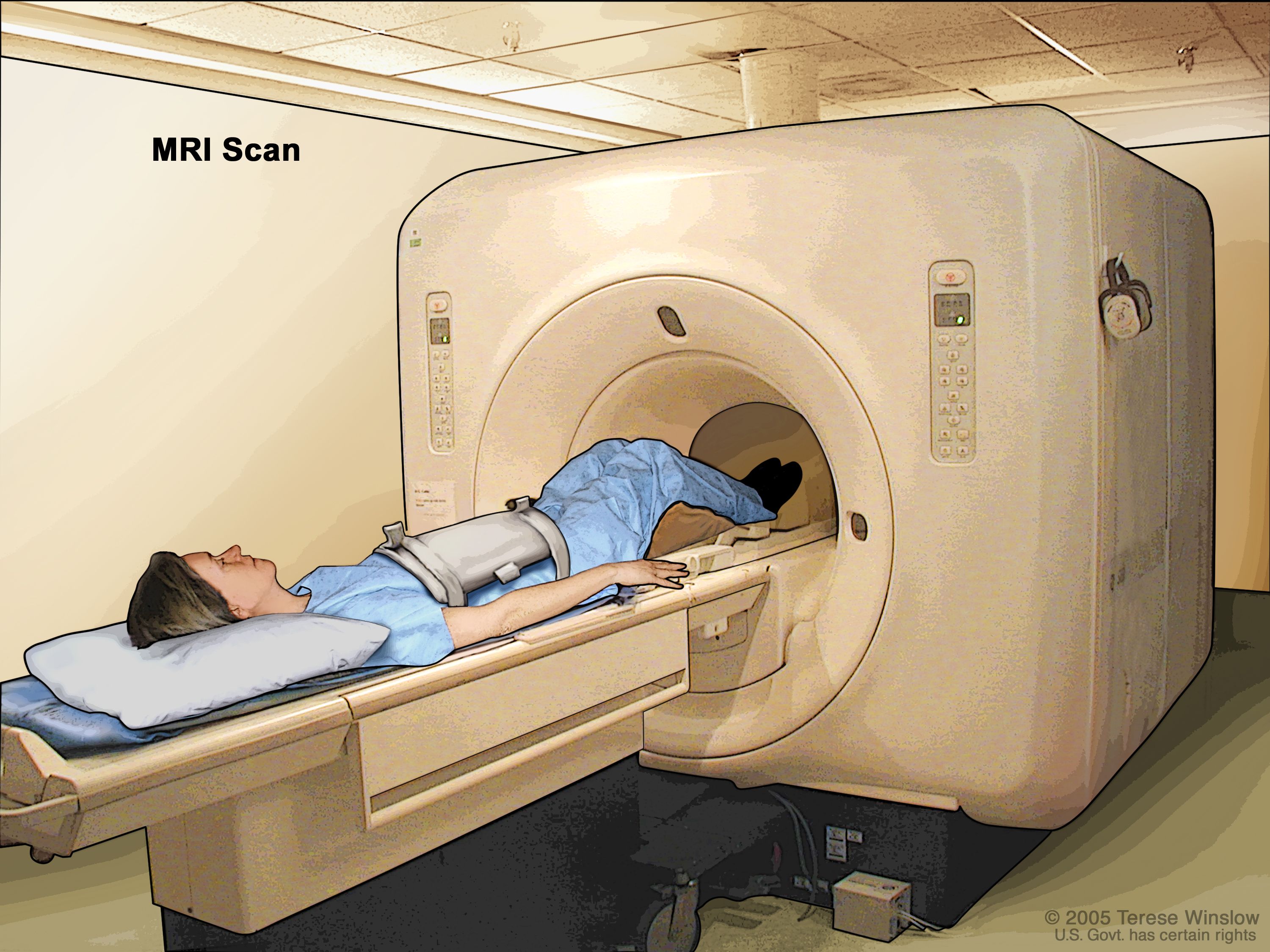
An MRI machine operates on principles that merge quantum physics with advanced computing. It starts with a large, powerful magnet that creates a temporary magnetic field around the patient’s body. Within our bodies, water molecules, which are abundant in living tissues, contain hydrogen atoms. These hydrogen atoms possess single protons that, under normal circumstances, spin randomly. However, when placed within the MRI’s strong magnetic field, these protons align themselves with the field, much like tiny compass needles orienting to the Earth’s magnetic north. This initial alignment is the first step in creating the raw material for our internal “photographs.”
Once aligned, a radiofrequency current is pulsed through the patient. This burst of radio waves temporarily knocks the aligned protons out of equilibrium, causing them to spin differently. When the radiofrequency pulse is turned off, the protons rapidly “relax” and realign themselves with the static magnetic field. As they do so, they release energy in the form of radio signals. The MRI machine’s highly sensitive antennae, acting like the advanced sensors in a high-resolution camera, detect these emitted signals.
The crucial aspect of this process, which allows for detailed imaging, is that different types of body tissues (e.g., bone, muscle, fat, diseased tissue) contain varying amounts of water and have distinct magnetic properties. Consequently, the time it takes for protons in these different tissues to realign, and the amount of energy they release, varies. A powerful computer then processes these subtle differences in emitted signals, converting them into a “digital image.” This transformation from raw signal to a comprehensible visual representation is akin to the “Converters” and “Optimizers” found on Tophinhanhdep.com, which take raw data and refine it into a polished, viewable image. The resultant images are cross-sectional “slices” of the body, offering a “three-dimensional” perspective that can be meticulously examined from multiple angles, providing invaluable diagnostic insights. The clarity and precision of these “digital images” are paramount, making the MRI an unparalleled tool for diagnosing complex medical conditions.
Varieties of Imaging: Open vs. Closed Systems, with and without Contrast
Just as Tophinhanhdep.com provides diverse “Image Collections” and “Photography” styles to suit different preferences, MRI technology offers variations designed to optimize both image quality and patient comfort. The two primary types of MRI machines are closed-bore and open-bore systems, each presenting distinct advantages.
Closed-Bore MRI Machines: These machines typically consist of a long, narrow tube where the patient lies. They are renowned for producing the “highest quality images” due to their stronger and more uniform magnetic fields. For many critical diagnoses where the utmost detail is required, such as in “Brain Tumors” or “Spinal Cord Conditions,” the closed-bore MRI is the preferred choice, offering a level of resolution comparable to the most pristine “High-Resolution Stock Photos” available on Tophinhanhdep.com. However, the enclosed nature of these machines can induce claustrophobia or anxiety in some patients, affecting their “emotional” comfort during the procedure.
Open-Bore MRI Machines: Developed to address concerns about claustrophobia and patient discomfort, open MRI machines feature two flat magnets positioned above and below the patient, leaving open space on two sides. This design significantly alleviates the feeling of being confined, making the experience more “Aesthetic” and tolerable for a broader range of individuals, including children and larger patients. While open MRIs enhance comfort, they generally produce images of slightly lower clarity compared to their closed-bore counterparts. This trade-off between comfort and peak “image quality” is a crucial consideration, much like choosing between a beautifully composed but lower-resolution “Aesthetic” background and a technically perfect, “High-Resolution” wallpaper on Tophinhanhdep.com.
MRI with Contrast Material: To further refine the “image quality” and enhance the visibility of specific structures or pathologies, some MRI exams incorporate contrast material. This contrast agent, often containing gadolinium, is injected intravenously. Once in the body, it alters the magnetic properties of nearby water molecules, causing them to realign more rapidly and emit stronger signals. The result is an “enhanced” image where areas like “Tumors,” “Inflammation,” “Infection,” and certain “Blood Vessels” appear brighter and more distinct. This technique is akin to using advanced “Editing Styles” or “Photo Manipulation” tools from Tophinhanhdep.com to highlight specific elements within an image, improving the “sensitivity and specificity” of the diagnostic “images.” While generally safe, the use of contrast agents requires careful consideration, particularly for pregnant individuals or those with severe kidney disease, underscoring the delicate balance between diagnostic clarity and patient safety.
A Comprehensive Visual Catalog: What MRI Scans Reveal
The true utility of MRI lies in its unparalleled ability to generate a diverse “Visual Catalog” of the body’s interior, allowing healthcare providers to diagnose a multitude of conditions with precision. Each scan contributes to an ever-growing “Image Inspiration & Collections” for medical professionals, offering “Photo Ideas” and “Thematic Collections” that inform crucial decisions about patient care. The versatility of MRI makes it an invaluable diagnostic tool across virtually every medical specialty.
From Brain to Bone: Unveiling Conditions Across Body Systems
MRI’s capacity to “unveil” conditions across the body’s various systems is a testament to its detailed imaging capabilities. It provides a unique “aesthetic” view into the human anatomy, transforming complex internal processes into interpretable “digital art.”
- Brain and Spinal Cord: For neurologists, MRI is the gold standard for “Brain and Neurological Conditions.” It can differentiate between white matter and gray matter, revealing subtle abnormalities that escape other imaging modalities. Conditions such as “Brain Aneurysms,” “Brain Tumors and Spinal Tumors,” “Multiple Sclerosis (MS)” lesions, and the effects of “Stroke” are visualized with exceptional clarity. It also helps in identifying injuries from “trauma” or issues like “Compression or inflammation of spinal cord and nerves” (pinched nerves). Each scan is a vital piece of the diagnostic puzzle, much like a carefully curated “Mood Board” provides a comprehensive visual overview for a creative project on Tophinhanhdep.com.
- Cardiac (Heart) Conditions: The heart, a dynamic and vital organ, benefits immensely from cardiac MRI. These “images” provide detailed insights into the “anatomy and function of your heart chambers, heart valves,” and blood flow through major vessels. This allows for the diagnosis of “Cardiovascular Conditions” such like “tumors, infections, and inflammatory conditions,” and the evaluation of “coronary artery disease.” For both children and adults with “congenital heart disease,” MRI offers an essential visual roadmap. The ability to capture the heart in motion, revealing its functional dynamics, is a form of advanced “digital photography” that informs complex surgical and treatment plans.
- Body (Chest, Abdomen, and Pelvis): When it comes to the torso, MRI offers a comprehensive view that often complements or surpasses other imaging techniques. It’s crucial for detecting “Tumors” in the chest, abdomen, or pelvis, diagnosing a range of “Liver Diseases” (e.g., cirrhosis, fatty liver, cancer), and assessing issues with the “biliary tract” and “pancreas.” “Inflammatory Bowel Disease” (Crohn’s disease, ulcerative colitis), “malformations of blood vessels,” and “inflammation of the vessels (vasculitis)” are also clearly depicted. Furthermore, MRI is safely used to monitor a “developing fetus in your uterus,” providing reassuring “visuals” for expectant parents. For female reproductive health, pelvic MRIs help diagnose “uterine fibroids, endometriosis, and ovarian cysts,” while in men, they are used to evaluate “prostate issues.”
- Bones and Joints: Athletes and individuals experiencing musculoskeletal issues frequently benefit from MRI. It’s unparalleled in evaluating “Bone Infections (osteomyelitis),” “Bone Tumors,” “Disk abnormalities in your spine,” and intricate “Joint Issues caused by injuries,” including “torn ligaments and muscles.” The detailed “photography” of soft tissues around bones, such as cartilage, ligaments, and tendons, makes MRI indispensable for sports medicine and orthopedics, offering “Beautiful Photography” of intricate anatomical structures.
- Breast Tissue: In conjunction with mammography, breast MRIs play a critical role in detecting “Breast Cancer,” particularly in individuals with “dense breast tissue” or those at “high risk.” These detailed “images” enhance the diagnostic process, helping to pinpoint suspicious areas with greater accuracy.
- Cancer Detection and Staging: Perhaps one of the most significant applications of MRI is in “Cancer Detection and Staging.” MRI’s “High-Resolution” capabilities allow clinicians to “identify both cancerous and non-cancerous tumors,” pinpoint their “exact location,” and measure their “size and spread.” This detailed “visual information” is crucial for staging cancer, guiding biopsies, and developing personalized treatment plans. Post-treatment, MRI is used for “Treatment Monitoring,” tracking whether tumors have shrunk, grown, or spread, creating a visual timeline of the disease’s progression. This application of MRI often deals with “Sad/Emotional” aspects, as the images carry profound implications for a patient’s life. However, they also offer “Inspiration” through the progress of treatment, akin to thematic “collections” on Tophinhanhdep.com that document a journey.
A Comparative Lens: MRI vs. CT in the Diagnostic Spectrum
Understanding “what is magnetic resonance imaging used for” often involves clarifying its relationship with other imaging technologies, particularly Computed Tomography (CT). While both are powerful diagnostic tools, they employ fundamentally different mechanisms and excel in different areas, much like distinct “Photography” techniques are chosen for different subjects.
MRI (Magnetic Resonance Imaging), as established, utilizes powerful magnets and radio waves to create detailed images. Its inherent strength lies in visualizing “non-bony parts or soft tissues” of the body. This includes structures like the “brain, spinal cord, nerves, muscles, ligaments, and tendons.” The images produced by MRI are renowned for their superior soft tissue contrast, allowing radiologists to distinguish between subtle tissue variations that might appear homogenous on a CT scan. This makes MRI the preferred choice for diagnosing conditions affecting the central nervous system, joints, and certain abdominal organs. A significant advantage of MRI is its safety profile; it “doesn’t use X-rays or other radiation,” making it a safer option for patients requiring repeated scans or for sensitive populations like pregnant women. This focus on non-invasiveness and patient safety is an “Aesthetic” value that sets MRI apart.
CT (Computed Tomography) scans, on the other hand, employ X-rays and computer processing to generate cross-sectional images. CT scans excel at visualizing “bony parts” of the body, as well as rapidly capturing images of organs, blood vessels, and soft tissues, particularly in emergency situations. CT is faster than MRI and is often the first choice for assessing trauma, detecting acute hemorrhages, or evaluating lung conditions. However, the use of X-rays means CT exposes patients to ionizing radiation, a factor that clinicians weigh carefully, especially for pediatric patients or those needing frequent imaging.
The decision between an MRI and a CT scan is a classic example of choosing the right “Image Tools” for the job, much like selecting specific “Converters” or “Compressors” on Tophinhanhdep.com based on the desired output. For example, if a doctor needs to see a fracture, a CT scan might be sufficient and quicker. But if they suspect a subtle ligament tear in a knee or an early brain tumor, the “High-Resolution” soft tissue contrast of an MRI provides the necessary detail, offering a “Beautiful Photography” of internal intricacies. While MRI is typically more expensive and can be contraindicated for patients with certain metal implants (e.g., pacemakers, some aneurysm clips) due to its strong magnetic field, these limitations are carefully considered against its diagnostic superiority for specific conditions. Ultimately, the choice between MRI and CT is guided by the clinical question, patient safety, and the need for optimal “image quality” to ensure an accurate diagnosis.
Beyond the Image: Safety, Preparation, and Patient Experience
While the primary function of MRI is to capture vital “images” for diagnostic purposes, the journey through an MRI scan involves a significant human element. Patient safety, meticulous preparation, and a thoughtful approach to the patient experience are paramount, addressing not just the physical but also the “Sad/Emotional” aspects that can arise during medical procedures. Just as Tophinhanhdep.com values the user’s interaction with visual content, the medical community prioritizes the patient’s interaction with the imaging process.
Ensuring Clarity and Comfort: Preparing for Your MRI
Proper preparation is crucial not only for patient safety but also for obtaining the highest “image quality” possible. The powerful magnetic field of an MRI scanner necessitates strict guidelines regarding metallic objects, which can interfere with the magnetic field, distort images, or even become dangerous projectiles.
- Metal and Electronics: Patients are instructed to remove all “Jewelry, watches, credit cards, hearing aids,” “pins, metal hair accessories, underwire bras, and metal zippers,” as well as “removable dental work,” “pens, pocketknives, eyeglasses,” “body piercings,” and “cell phones, electronic watches, and tracking devices.” This is akin to a photographer preparing their studio, ensuring all elements that could cause glare or distortion are removed to capture a perfect “digital photography.”
- Implanted Devices: A comprehensive medical history is taken to identify any “implanted medical devices” such as “heart pacemakers or defibrillators, electronic or implanted stimulators (deep brain, vagus nerve, bladder, spine, neurostimulators), metallic joint prostheses, cochlear implants,” or “aneurysm clips and coils.” Many newer implants are MRI-safe, but others are not, requiring careful screening to prevent malfunction or injury. This pre-screening is a critical step in “Visual Design,” ensuring the canvas is clear and safe for the imaging process.
- Personal Considerations: Patients are also asked about “pregnancy” (as gadolinium contrast is generally avoided), the ability to “lie on your back for 30 to 60 minutes,” and any history of “claustrophobia.” For those with a fear of enclosed spaces, options like “sedatives” or even “anesthesia” are discussed, and “open MRI” machines are considered where appropriate. This proactive approach to patient comfort acknowledges the “emotional” aspect of undergoing a scan, ensuring the experience is as stress-free as possible.
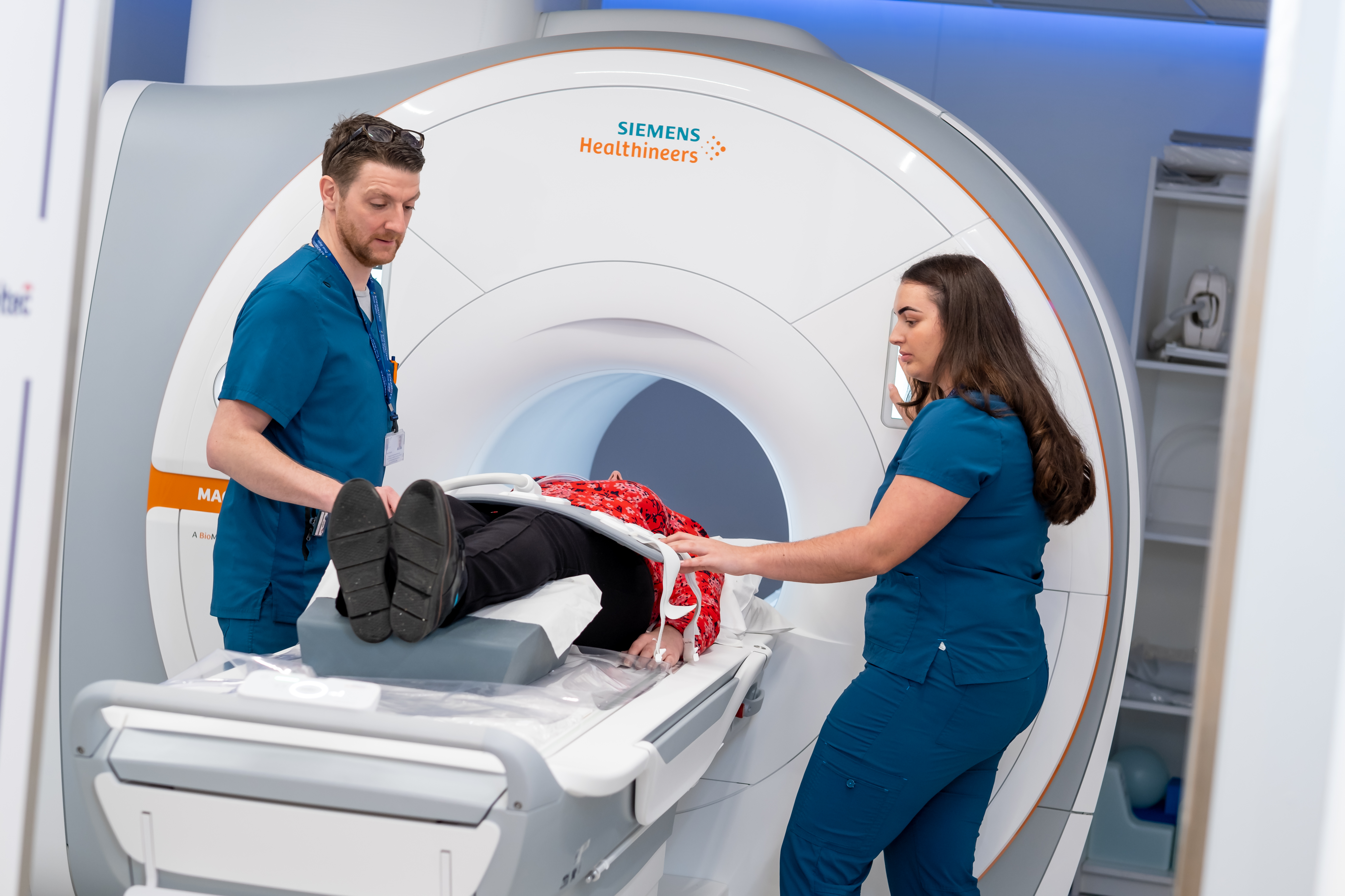
During the scan itself, patients are often given earplugs or headphones to mitigate the “loud knocking and clicking sounds” generated by the machine. The importance of “being very still during the exam” is emphasized to ensure optimal “image quality,” preventing blurriness that would render the “photography” diagnostically useless. Communication is maintained through an intercom system, and a call button allows patients to alert the technologist to any issues, fostering a sense of control and safety.
Navigating the Nuances: Safety and Considerations for MRI
While MRI is generally considered “safe,” particularly when compared to radiation-emitting technologies, there are important “nuances” and considerations that healthcare providers carefully manage to optimize patient outcomes. These safety protocols are integral to the ethical “Visual Design” of medical diagnostics.
- Magnetic Field Safety: The “strong magnetic field” itself is not harmful to the patient, but its powerful force on ferromagnetic objects requires strict adherence to safety guidelines. Uncontrolled metallic objects can cause serious injury. This risk assessment is a foundational aspect of MRI safety, ensuring that the environment for capturing “inner images” is secure.
- Contrast Material Risks: While “contrast materials are safe drugs” for most, there’s a “very slight risk of an allergic reaction,” which is usually mild and manageable. More significantly, “Nephrogenic Systemic Fibrosis (NSF)” is a “rare but serious complication” in individuals with “severe kidney disease” who receive gadolinium-based contrast. Therefore, use in these patients is carefully evaluated, and dialysis may be recommended promptly after the scan to remove the agent. There’s also emerging evidence of “tiny traces of gadolinium” potentially remaining in organs after contrast-enhanced MRI, though “no known negative effects” from this have been definitively established. This ongoing research reflects a commitment to continuous improvement in imaging safety.
- Pregnancy and Fetal MRI: Healthcare providers generally “don’t perform gadolinium contrast-enhanced MRIs on pregnant women” unless “absolutely necessary,” due to unknown risks to the developing fetus, especially during the crucial first trimester when organs are forming. However, “Fetal MRI” without contrast is increasingly used to evaluate a developing fetus for abnormalities detected on ultrasound, offering highly detailed “images” that guide prenatal care. This demonstrates a careful balance between the diagnostic benefits of “Beautiful Photography” and the paramount importance of maternal and fetal safety.
- Claustrophobia Management: The “claustrophobia” experienced by some patients in closed MRI machines is a significant “emotional” challenge. Beyond sedatives and open MRI options, facilities may offer “coping mechanisms” such as listening to music, watching videos, or using visualization techniques. The goal is to make the experience less daunting, ensuring that the patient can remain still for the required duration, which directly impacts the “High Resolution” of the diagnostic “images.” These accommodations are part of a patient-centered approach to medical “Visual Design,” prioritizing both clinical effectiveness and individual well-being.
In summary, the sophisticated process of Magnetic Resonance Imaging extends far beyond merely generating “images.” It encompasses a rigorous system of safety protocols, detailed patient preparation, and a compassionate approach to patient comfort. This holistic perspective ensures that MRI remains a trusted and invaluable diagnostic tool, consistently delivering “High Resolution” “digital photography” of the body’s interior, which in turn leads to clearer diagnoses and more effective treatment pathways, mirroring the precision and care evident in the “Photography” and “Image Tools” found on Tophinhanhdep.com.
The journey from initial symptoms to a clear diagnosis often hinges on the ability to see what lies beneath the surface. Magnetic Resonance Imaging, with its capacity to render the body’s hidden world in exquisite “High Resolution” detail, stands as a beacon of modern medical technology. Just as Tophinhanhdep.com empowers individuals to explore, create, and appreciate the vast spectrum of visual art, MRI empowers healthcare providers to uncover, understand, and address the intricate “visual designs” of human health and disease.
From identifying the subtle signs of “Brain Tumors” to charting the course of “Heart Disease” or pinpointing the exact location of “Torn Ligaments,” MRI provides a “Comprehensive Visual Catalog” that is unrivaled. Its ability to achieve this without the use of ionizing radiation underscores a commitment to patient safety, making it a preferred choice for numerous diagnostic challenges. The evolution of MRI, including open-bore designs and advanced contrast techniques, continually refines its role, balancing diagnostic power with patient comfort—a true “aesthetic” consideration in medical practice.
Ultimately, “what is magnetic resonance imaging used for” transcends a simple list of medical applications. It represents a profound convergence of science, technology, and compassionate care. It’s a testament to humanity’s ongoing quest to understand itself, using sophisticated “Image Tools” to capture the most intimate “Photography” of life itself. In every scan, MRI creates an invaluable piece of “Digital Art,” a diagnostic masterpiece that guides millions towards better health, reinforcing the vital role that clear, detailed images play, not just in our digital lives through platforms like Tophinhanhdep.com, but critically, within the realm of saving and improving lives. If you or a loved one are facing unexplained symptoms, consult with your healthcare provider about whether an MRI could illuminate the path to diagnosis and recovery, offering the clarity of vision needed for a brighter, healthier future.