Decoding the Invisible: What is Medical Imaging and Its Role in Modern Healthcare

In an era where visual information reigns supreme, from breathtaking Beautiful Photography gracing Wallpapers to intricate Digital Art inspiring Creative Ideas on platforms like Tophinhanhdep.com, the realm of medical imaging stands as a vital, often life-saving, frontier of visual exploration. Medical imaging is, at its core, the intricate process of creating visual representations of the interior of the human body. These aren’t merely Stock Photos; they are high-stakes, High Resolution visual records that allow medical professionals to peer beyond the skin and bones, revealing the delicate machinery and intricate biology that define human health.
The significance of medical imaging in today’s healthcare system cannot be overstated. It acts as the eyes of modern medicine, empowering doctors and allied health professionals to diagnose diseases, monitor treatment efficacy, and guide surgical interventions with unparalleled precision. Just as a photographer employs specific lenses and Editing Styles to capture the perfect shot, medical imaging specialists utilize a diverse array of advanced technologies and Image Tools to capture the clearest, most diagnostic images possible. These images, often presented in formats far more complex than simple Backgrounds, form the foundation of countless medical decisions, impacting millions of lives globally.
This comprehensive guide delves into the essence of medical imaging, exploring its diverse modalities, the pivotal roles of the professionals who operate these sophisticated systems, and the dynamic educational pathways leading to a rewarding career in this ever-evolving field. We will also touch upon how the principles of image clarity, visual design, and data processing, fundamental to Tophinhanhdep.com’s mission, find profound real-world application in the critical domain of medical diagnostics.
The Core of Medical Imaging: Visualizing the Unseen
At its heart, medical imaging is about making the invisible visible. It’s about capturing a moment, a structure, or a physiological process within the body that cannot be perceived by the naked eye. This capability is paramount for early disease detection, accurate diagnosis, and effective treatment planning. Without these internal “photographs,” much of modern medicine would be relegated to guesswork, leading to less effective and often more invasive interventions.
The field has seen remarkable growth since its inception, continually pushing the boundaries of what can be seen and understood about the human body. Early medical imaging was rudimentary, a basic “snapshot” compared to today’s vibrant, multi-dimensional views. Today, the technology involved is nothing short of revolutionary, offering a spectrum of tools, each with its unique strengths, much like a graphic designer choosing the right software for a specific Visual Design project. The evolution from flat, static images to dynamic, interactive 3D and even 4D representations mirrors the advancements seen in digital photography and Digital Art, where static prints have given way to immersive visual experiences.
The core purpose of medical imaging can be broken down into three primary objectives:
- Diagnosis: Identifying the presence, nature, and extent of a disease or injury.
- Monitoring: Tracking the progression of a condition or the effectiveness of a treatment over time.
- Treatment Guidance: Providing real-time visual information to guide minimally invasive procedures or surgeries.
From a Tophinhanhdep.com perspective, these images are specialized forms of “photography” – not of nature’s landscapes or abstract patterns, but of the intricate biological landscape within us. Each scan, whether an X-ray of a broken bone or an MRI of brain activity, captures a critical “photo idea” for medical analysis.
A Brief History and Evolution
The journey of medical imaging began in 1895 with Wilhelm Conrad Roentgen’s groundbreaking discovery of the X-ray. This accidental revelation allowed the first glimpse into the living human body, laying the cornerstone for all subsequent advancements. What started as a simple, almost Abstract shadow image, quickly became a foundational diagnostic tool.
The early 20th century saw the development of various X-ray-based techniques, expanding Roentgen’s initial discovery. Mammography, specifically designed for breast tissue imaging, emerged in the 1920s, followed by tomography (the precursor to modern CT) in the 1930s, angiography (for blood vessels) in the 1940s, and fluoroscopy (real-time X-ray imaging) in the 1950s. These developments were akin to photographers discovering new film types and developing techniques, each offering a clearer, more nuanced “picture.”
The 1950s also marked a significant leap with the advent of nuclear medicine, which used radioactive tracers to visualize physiological processes rather than just anatomy. This led to the development of Positron Emission Tomography (PET) scans, revolutionizing cancer diagnosis and metastasis detection.
The 1970s brought another wave of transformative technologies: ultrasound, Computed Tomography (CT), and Magnetic Resonance Imaging (MRI). Ultrasound’s ability to image soft tissues and, notably, the brain (a previously challenging area) without radiation was a major breakthrough. CT scans provided detailed cross-sectional views, and MRI offered unprecedented detail of soft tissues and functional information, even allowing real-time monitoring of treatments.
Today, medical imaging is at an exciting crossroads, driven by rapid technological advancements. Techniques such as 3D and 4D imaging are becoming standard, offering dynamic, volumetric data that allows doctors to visualize complex structures and processes with incredible clarity, much like AI Upscalers enhance image resolution on Tophinhanhdep.com. The integration of Artificial Intelligence is also transforming image acquisition, processing, and interpretation, promising earlier, less invasive detection of diseases, aligning with the “smart tools” philosophy of modern digital image management.
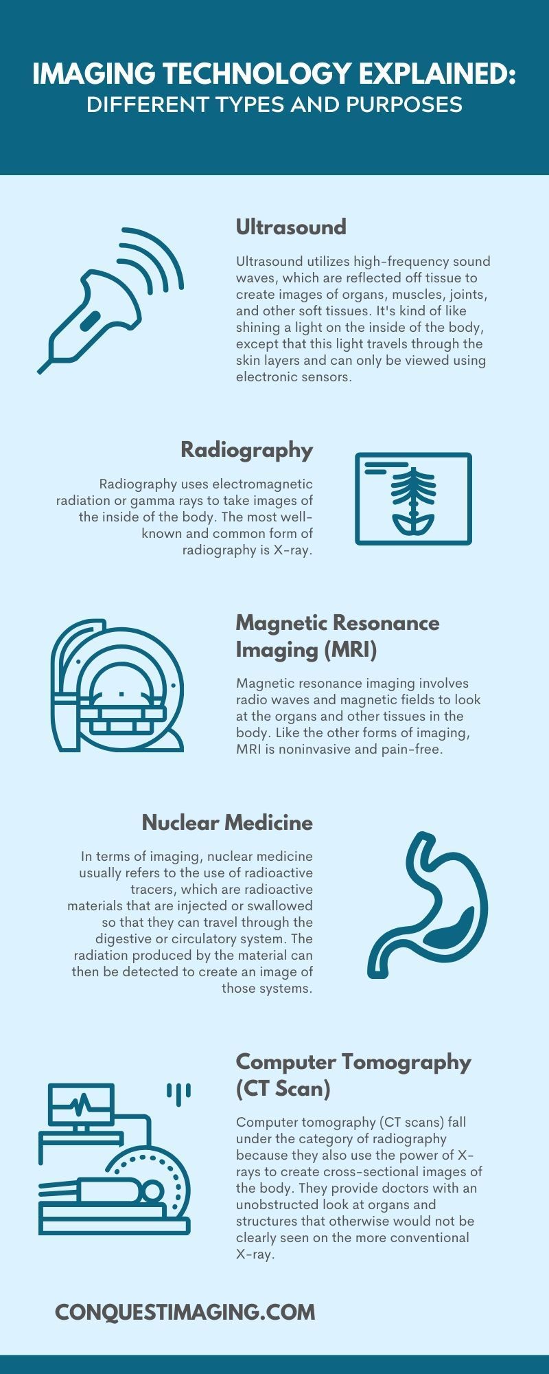
Specialized Imaging Modalities: A Spectrum of Diagnostic Power
Just as Tophinhanhdep.com offers diverse categories from Nature Photography to Sad/Emotional images, medical imaging employs various modalities, each tailored to specific diagnostic needs and biological structures. Understanding these different technologies is crucial for appreciating the depth and breadth of this field.
Computed Tomography (CT)
Computed Tomography, often known as a CT scan or CAT scan (Computerized Axial Tomography), uses X-ray radiation to create detailed cross-sectional images of the body. Unlike a traditional X-ray, which produces a single 2D image, a CT scanner rotates around the patient, taking numerous images from different angles. A computer then processes these images to generate highly detailed slices, or “tomograms,” of bones, soft tissues, and blood vessels.
From a visual perspective, CT images can be thought of as a series of intricate digital photographs, each revealing a thin layer of the body’s internal architecture. The data generated is immense, requiring robust Image Tools for storage, retrieval, and viewing. These scans are invaluable for diagnosing internal injuries after trauma, pinpointing tumors, detecting infections, and monitoring the progression of diseases like cancer or heart conditions. The ability to render these slices into 3D models further enhances their diagnostic utility, akin to creating Digital Art from raw photographic data.
Magnetic Resonance Imaging (MRI)
Magnetic Resonance Imaging (MRI) stands apart by using powerful magnetic fields and radio waves, rather than ionizing radiation, to generate images. The patient lies within a large, tunnel-like magnet, which aligns the protons within the body’s water molecules. Radiofrequency currents are then pulsed through the patient, briefly knocking these protons out of alignment. When the radiofrequency is turned off, the protons relax back into alignment, emitting energy that is detected by the scanner and converted into detailed images.
MRI excels at visualizing soft tissues such as the brain, spinal cord, nerves, muscles, ligaments, and tendons. It’s an indispensable tool for diagnosing strokes, tumors, aneurysms, eye and inner ear disorders, and various musculoskeletal injuries. For organ imaging, it can assess heart function, the effects of a heart attack, blood vessel inflammation, and detect tumors in organs like the liver, breast, ovaries, kidneys, and pancreas. The aesthetic quality of an MRI, often presented in grayscale with distinct tissue contrast, offers a unique, almost Abstract view of the body’s internal landscape, capturing the subtle nuances that define health and disease. This modality emphasizes High Resolution to resolve minute structural details.
Ultrasound Imaging (Sonography)
Ultrasound imaging, or sonography, utilizes high-frequency sound waves to create real-time moving images of internal structures. A sonographer applies a gel to the skin, then uses a handheld device called a transducer. The transducer emits sound waves into the body, which bounce off organs and tissues and return to the transducer. These reflected sound waves are then converted into electrical signals, which a computer transforms into a live, dynamic image on a screen.
The absence of radiation makes ultrasound particularly safe and ideal for monitoring fetal growth and development during pregnancy, a truly Beautiful Photography moment within the body. It’s also widely used to diagnose conditions related to pain, swelling, or infection in various organs, including the heart, gallbladder, liver, kidneys, brain (in infants), and spine. The real-time nature of ultrasound images means it captures not just static snapshots, but dynamic physiological processes, offering a form of live “digital photography” of the internal environment. The portability of ultrasound equipment also makes it invaluable in emergency situations, allowing quick bedside diagnostics.
X-ray and Fluoroscopy
Conventional X-ray radiography, the oldest form of medical imaging, uses small doses of ionizing radiation to produce static images of the body’s internal structures. X-rays pass through the body, and different tissues absorb the radiation at varying rates; bones, for example, appear white because they absorb a lot of radiation, while air-filled lungs appear black.
Fluoroscopy is a specialized X-ray technique that produces a continuous X-ray image, much like a movie. This “live-action” X-ray allows doctors to observe the movement of organs, visualize blood flow, or guide catheters during procedures. While relatively simple compared to CT or MRI, X-rays remain a primary tool for detecting bone fractures, pneumonia, and foreign objects, offering quick, fundamental “background” information for diagnostics. The simplicity and speed often make X-rays the first line of visual defense in emergency medicine.
Nuclear Medicine and Molecular Imaging
Nuclear medicine employs small amounts of radioactive materials, called radiotracers, to visualize organ function and molecular processes within the body. Unlike other modalities that primarily show anatomy, nuclear medicine focuses on physiology. For example, a PET scan can detect metabolic activity, which is crucial for identifying cancerous cells, tracking disease spread, or assessing brain function.
These images often appear more Abstract than anatomical scans, showing areas of varying metabolic activity as glowing regions. They provide unique “image inspiration” for understanding disease at a cellular level, allowing doctors to detect diseases like cancer, heart disease, and neurological disorders at their earliest stages. The combination of structural imaging (like CT or MRI) with functional imaging (like PET) creates a powerful hybrid, offering a comprehensive “photo idea” of both form and function.
Advanced Mammography Techniques
Mammography, a specific type of X-ray imaging for the breast, has also undergone significant advancements. While traditional mammography provided 2D film images, modern digital mammography offers clearer images with lower radiation doses. Furthermore, 3D mammography (breast tomosynthesis) creates a layered image of the breast, which can detect smaller cancers and reduce false positives, especially in women with dense breast tissue. These detailed, multi-layered “photographs” are critical for early breast cancer detection, offering a more complete “visual design” of breast health.
The Human Element: Diverse Careers in Medical Imaging
The sophisticated technologies of medical imaging are only as effective as the skilled professionals who operate them and interpret their output. Just as Tophinhanhdep.com relies on talented photographers and visual designers, the medical imaging field relies on a dedicated team of experts. These careers offer significant job security, competitive salaries, and the profound satisfaction of directly impacting patient care, making a real difference beyond creating Aesthetic images.
The Medical Imaging Team
A diverse team works together to ensure accurate and safe imaging procedures:
- Radiologic Technologists (Radiographers): These professionals are at the forefront of image acquisition, primarily operating X-ray, CT, and sometimes MRI equipment. They are responsible for patient preparation, positioning, operating the machinery, and ensuring image quality, akin to a professional photographer setting up a shot.
- Diagnostic Medical Sonographers (Ultrasound Technicians): Specializing in ultrasound, sonographers skillfully manipulate transducers to capture dynamic images of soft tissues, fetal development, and blood flow. Their work requires excellent hand-eye coordination and the ability to interpret real-time visuals.
- MRI Technologists: Experts in magnetic resonance imaging, these technologists operate MRI scanners, ensuring patient safety (especially regarding metal objects), preparing patients, and producing high-quality images of internal structures.
- Nuclear Medicine Technologists: These specialists administer radiotracers and operate nuclear medicine cameras (like PET and SPECT scanners) to capture images of organ function, playing a critical role in diagnosing and monitoring physiological processes.
- Vascular Interventional Radiographers: Working closely with interventional radiologists, these professionals assist in minimally invasive, image-guided procedures, primarily using fluoroscopy and other imaging modalities to navigate catheters and instruments within the body.
- Radiation Therapists: While not directly involved in diagnostic imaging, radiation therapists use imaging technologies (often CT scans) to precisely plan and deliver radiation treatment for cancer patients.
- Radiologists: These are licensed medical doctors with extensive advanced training in medical imaging and diagnostics. Their main responsibility is to accurately interpret the images generated by technologists, provide detailed reports, and consult with referring physicians and patients. They are the “art critics” or “editors-in-chief” of the medical imaging world, turning raw visual data into actionable diagnoses.
- Radiologic Nurses: Registered Nurses who specialize in caring for patients undergoing medical imaging procedures, particularly those involving contrast media or sedation.
Essential Skills and Personality Traits
A career in medical imaging demands a unique blend of technical proficiency and interpersonal skills. Professionals in this field often possess traits that would serve well in any detail-oriented, visually-driven profession:
- Organized and Detail-Oriented: Crucial for precise patient positioning, equipment calibration, and meticulous record-keeping.
- A People Person with Excellent Communication Skills: Interacting with anxious patients requires empathy, clear explanation, and the ability to reassure.
- Level-Headed and Patient: Maintaining composure in high-pressure situations and during lengthy procedures is key.
- Good Hand-Eye Coordination and Concentration: Essential for operating complex equipment and capturing optimal images, much like a photographer needing steady hands for the perfect shot.
- Dependable: Healthcare settings demand reliability and consistency.
These individuals are not just operating machines; they are artists and scientists in one, capturing the intricate “nature” of human health with precision and care.
Beyond Diagnosis: The Broader Impact of Medical Imaging
The utility of medical imaging extends far beyond simply diagnosing an ailment. It forms a cornerstone of preventive medicine, guides intricate therapeutic interventions, fuels medical research, and plays a critical role in public health education. From the perspective of Tophinhanhdep.com, these applications highlight the vast Image Inspiration & Collections that medical visuals provide, driving continuous innovation and understanding.
Prevention and Screening
One of the most powerful applications of medical imaging is in preventive screening. Detecting diseases in their earliest, most treatable stages can dramatically improve patient outcomes and even save lives. Mammograms for breast cancer, CT lung screenings for high-risk individuals, and specialized cardiac imaging to assess heart disease risk are prime examples.
These screenings are a form of proactive “photography,” capturing potential issues before they manifest as severe symptoms. The data gathered from these wide-scale screenings also provides invaluable Thematic Collections of images for medical research, aiding in the development of new technologies and improved treatment protocols. By informing the public about the benefits of early detection, healthcare professionals leverage visual evidence to improve general knowledge and dispel common misconceptions about medical procedures.
Treatment and Therapy Guidance
Medical imaging is not just for diagnosis; it’s an indispensable guide for many modern treatments. Interventional radiology, for instance, uses real-time imaging (like fluoroscopy, CT, or ultrasound) to guide minimally invasive procedures. This allows specialists to perform angioplasties, place stents, remove blockages, or biopsy tumors through tiny incisions, reducing patient discomfort, recovery time, and costs.
Therapeutic radiology, or radiation therapy, also heavily relies on imaging. CT scans are used to precisely map the location of a tumor, enabling radiation therapists to plan and deliver highly targeted radiation doses while sparing surrounding healthy tissues. This precise “photo manipulation” of radiation beams ensures maximum efficacy with minimal side effects. The imaging process is fundamental to ensuring that the “creative ideas” for treatment are executed with pinpoint accuracy.
Tracking and Evaluation of Treatment Response
Following a diagnosis and the initiation of treatment, medical imaging becomes a critical tool for monitoring progress. Serial scans provide objective, reproducible results that clearly show changes in a patient’s condition over time and measure their specific response to therapy. Is a tumor shrinking? Is an infection clearing? Is a fracture healing correctly? Imaging provides the definitive answers.
This continuous visual feedback helps clinicians determine if a chosen treatment plan is working or if adjustments are needed. For chronic diseases like cancer, regular radiological studies are essential for long-term surveillance, offering a visual “timeline” of the patient’s health journey. This creates an ongoing “collection” of images that tell a powerful story, much like a photographic series depicting a significant life event.
Driving Research and Technological Advancement
The medical imaging field is constantly evolving, with new technologies and applications emerging regularly. Radiologists, physicists, and biomedical engineers continually engage in research, using current imaging capabilities to explore new surgical procedures, develop innovative medical devices, and refine treatment methods.
Platforms like Tophinhanhdep.com showcase Trending Styles in visual arts; similarly, medical imaging research highlights the “trending styles” in diagnostic innovation. From AI Upscalers that enhance image clarity to advanced algorithms that automate anomaly detection, the integration of artificial intelligence is rapidly transforming how medical images are acquired, processed, and interpreted. These innovations aim to improve diagnostic accuracy, reduce scan times, and make imaging more accessible, embodying the “Image Tools” philosophy of continuous improvement.
Crafting a Future in Medical Imaging: Education, Certification, and Professional Growth
For those drawn to the precision of Digital Photography and the profound impact of visual information, a career in medical imaging offers a dynamic and rewarding path. The demand for skilled professionals in this field is consistently high, driven by an aging global population and continuous advancements in diagnostic technology. Aspiring imaging professionals must navigate specific educational and certification requirements, ensuring they are well-prepared to contribute to this vital sector of healthcare.
Educational Pathways and Accreditation
Entering the medical imaging field typically involves one of several educational paths, ranging from certificates to bachelor’s degrees. The choice often depends on the desired specialization and career advancement goals.
- Certificate Programs: Often suitable for those already in healthcare, offering specialized training in a medical imaging modality in about six months to a year.
- Associate’s Degree: A common pathway for many roles, such as radiologic technologists and diagnostic medical sonographers, typically taking two years. These programs often combine classroom instruction with extensive clinical education.
- Bachelor’s Degree: Provides more in-depth instruction and a broader education, preparing individuals for technologist roles, management positions, or further graduate studies, essential for those aspiring to become radiologists.
Regardless of the chosen path, selecting an accredited school or program is paramount. Accreditation ensures that the education meets recognized standards of quality and prepares graduates for professional certification and licensure. Key accrediting bodies include:
- Commission on Accreditation for Allied Health Education Programs (CAAHEP): Collaborates with specialized review committees (e.g., JRC-DMS for Diagnostic Medical Sonography, JRCCVT for Cardiovascular Technology).
- Joint Review Committee on Education in Radiologic Technology (JRCERT): Accredit programs for radiologic technology.
- Joint Review Committee on Education Programs in Nuclear Medicine Technology (JRCNMT): Accredit programs for nuclear medicine technology.
Accreditation not only guarantees a quality education but also often makes graduates eligible for federal financial aid and, crucially, for professional certification exams.
Core Coursework
A solid foundation in science and human anatomy is fundamental for any medical imaging professional. Common coursework across various programs includes:
- Anatomy and Physiology: Understanding the structure and function of the human body.
- Pathophysiology: Studying the mechanisms of disease.
- Physics: Comprehending the principles behind imaging technologies (e.g., X-ray physics, ultrasound physics, magnetic resonance physics).
- Medical Terminology: Essential for effective communication in healthcare.
- Radiologic Science/Ultrasound Scanning/Ultrasound Instrumentation: Specific technical training related to the chosen modality.
- Clinical Education: Hands-on experience in real healthcare settings, applying theoretical knowledge under supervision.
These rigorous academic and practical components ensure that graduates are not only technically proficient but also possess a deep understanding of the medical context in which their “images” are created and interpreted.
Professional Certification and Licensure
Most employers prefer, and many states require, medical imaging professionals to hold professional certification and/or licensure. Certification demonstrates competency and adherence to professional standards.
- American Registry of Radiologic Technologists (ARRT): The primary certification body for radiologic technologists, MRI technologists, and radiation therapists. Certification typically requires graduating from an accredited program and passing a comprehensive exam.
- American Registry for Diagnostic Medical Sonography (ARDMS): Certifies sonographers, often in various specialties such as abdominal, obstetric/gynecologic, or vascular sonography.
- Nuclear Medicine Technology Certification Board (NMTCB): Certifies nuclear medicine technologists.
- Cardiovascular Credentialing International (CCI): Offers certifications for cardiovascular technologists.
State-specific licensing requirements vary, so it is essential for aspiring professionals to consult their state’s board of health for specific mandates. Obtaining multiple credentials or specialized certifications can significantly enhance career prospects and earning potential, much like a photographer diversifying their portfolio with Nature or Aesthetic shots.
Job Growth and Salary Prospects
The outlook for medical imaging careers is exceptionally strong, largely due to an aging population that requires increasing healthcare services and the continuous evolution of imaging technologies. The U.S. Bureau of Labor Statistics projects robust growth across the field:
- Diagnostic Medical Sonographers: Anticipated growth rate of 15.1% through 2033, significantly faster than the national average for all occupations. Median annual salary around $89,340.
- Radiologic and MRI Technologists: Projected job growth rate of 5.8% through 2033, also ahead of the national average. Median annual salaries are around $73,410 for radiologic technologists and $88,180 for MRI technologists.
- Radiation Therapists: Expected growth of approximately 3.1%. Median annual salary around $101,990.
- Cardiovascular Technologists: Projected growth of 10% by 2032, with median salaries around $63,000 to $81,000+.
These figures underscore the stability and financial rewards associated with medical imaging careers, offering a compelling alternative to roles that might solely focus on creating Wallpapers or Backgrounds.
Professional Networking and Continuous Learning
In a field as dynamic as medical imaging, continuous learning and professional networking are vital for staying relevant. Joining professional organizations offers invaluable opportunities for networking, accessing continuing education, and staying abreast of technological advancements and best practices. Notable organizations include:
- The Society of Diagnostic Medical Sonography (SDMS)
- American Registry of Radiologic Technologists (ARRT)
- Society of Nuclear Medicine and Molecular Imaging (SNMMI)
- Cardiovascular Credentialing International (CCI)
- American Society of Radiologic Technologists (ASRT)
These organizations serve as “mood boards” of professional development and “thematic collections” of knowledge, ensuring that professionals can continually hone their “visual design” and technical skills.
Conclusion
Medical imaging is a fascinating and indispensable component of modern healthcare, transforming our ability to see, diagnose, and treat conditions within the human body. It is a field that perfectly marries cutting-edge technology with the critical need for human expertise, where precision, visual acuity, and compassionate patient care are paramount.
From the discovery of the X-ray to today’s multi-modal, AI-enhanced systems, medical imaging continues to push the boundaries of what’s possible, much like the continuous innovation seen in Digital Photography and Visual Design. For those with a passion for technology, a keen eye for detail, and a desire to make a tangible difference in people’s lives, a career in medical imaging offers a uniquely rewarding path.
Just as Tophinhanhdep.com provides a platform for Image Inspiration & Collections, medical imaging provides a profound source of inspiration for understanding life itself. The images captured by these dedicated professionals are far more than mere visuals; they are windows into health, hope, and the human condition, forming an irreplaceable “background” to the intricate tapestry of modern medicine.