Exploring the Inner Canvas: What Magnetic Resonance Imaging Reveals

The human body is an intricate masterpiece, a complex tapestry of tissues, organs, and systems working in perfect, often hidden, harmony. For centuries, understanding its internal workings required invasive procedures, but advancements in medical technology have revolutionized our ability to peer inside without a single incision. Among these innovations, Magnetic Resonance Imaging (MRI) stands out as a triumph of physics and engineering, offering unparalleled insights into the body’s internal architecture. At Tophinhanhdep.com, we celebrate the power of visuals, from stunning wallpapers and aesthetic photography to sophisticated digital art and image tools. In that spirit, we delve into the world of MRI, a technology that crafts high-resolution “images” of our biological essence, transforming unseen complexities into clear, diagnostic “photography.”

MRI is far more than just a diagnostic scan; it is a sophisticated “image tool” that creates detailed, three-dimensional “visual designs” of anatomy and physiological processes. Unlike X-rays or CT scans, which rely on ionizing radiation, MRI harnesses powerful magnetic fields and radio waves, making it a safer option for repeated examinations. This non-invasive nature and its exceptional ability to differentiate between various types of soft tissues have made MRI an indispensable resource in modern medicine. Just as a skilled photographer captures the nuanced beauty of nature or an abstract concept, an MRI machine “photographs” the subtle distinctions within our bodies, providing medical professionals with critical “image inspiration” for accurate diagnosis, effective treatment planning, and continuous monitoring of health conditions.
Understanding Magnetic Resonance Imaging: A Deeper Look into its Visual Power
To truly appreciate MRI, we must first understand the remarkable science that underpins its ability to generate such breathtakingly detailed internal “images.” It’s a process that, in essence, turns the human body into a canvas upon which intricate physiological patterns are revealed through electromagnetic interactions.
The Science Behind the Image: How MRI Works
At its core, an MRI scan functions by leveraging the magnetic properties of hydrogen atoms, which are abundant in the water molecules found throughout living tissues. When a patient is placed inside the large, powerful magnet of an MRI machine, these hydrogen protons, which naturally spin and wobble, align themselves with the direction of this strong magnetic field. Think of it like aligning tiny compass needles in a giant, unseen force field.
Next, a radiofrequency current is briefly pulsed through the patient. This burst of energy momentarily “excites” the aligned protons, causing them to tip out of alignment and spin out of equilibrium, momentarily straining against the pull of the main magnetic field. When this radiofrequency pulse is turned off, the protons relax and rapidly “realign” with the powerful magnetic field. As they realign, they release energy, which is detected by sophisticated sensors within the MRI scanner.
The magic of MRI lies in the subtle differences in this realignment process. The speed at which protons realign and the amount of energy they release vary significantly depending on their surrounding environment and the chemical nature of the molecules they are part of. For instance, protons in fatty tissue will realign differently than those in water-rich organs like the brain or muscles. The MRI computer processes these varying signals, converting them into a digital “high-resolution” image, much like how a digital camera captures light variations to form a photograph. This intricate process allows radiologists to distinguish between different tissue types—healthy, diseased, or injured—based on their unique magnetic properties, essentially creating a “digital photography” of the body’s interior.
Sometimes, to enhance the clarity and detail of these internal “images,” a contrast agent, often containing the element Gadolinium, may be administered intravenously before or during the scan. This agent temporarily alters the magnetic properties of tissues, causing protons in certain areas to realign more quickly, resulting in brighter “photography” in those specific regions. This is akin to applying an “editing style” in post-production to highlight particular features in a photograph, making abnormalities like tumors or inflammation stand out with greater prominence. Just as a photographer requires a steady hand for sharp images, patients must remain very still during an MRI to prevent blurring and ensure the capture of truly “high-resolution” diagnostic visuals.
MRI vs. Other Imaging Techniques: A Visual Comparison
In the realm of medical imaging, MRI is often compared with other common diagnostic tools such as X-rays and Computed Tomography (CT) scans. While all aim to visualize the body’s interior, their underlying principles and applications differ significantly, akin to different genres or “editing styles” in the world of “photography” available on Tophinhanhdep.com.
The most fundamental distinction lies in their energy source. X-rays and CT scans utilize ionizing radiation, which, while beneficial for diagnosis, carries a small risk of cellular damage with repeated exposure. MRI, however, entirely avoids ionizing radiation, relying instead on magnetic fields and radio waves. This crucial safety advantage makes MRI the “image tool of choice” when frequent imaging is required, particularly for vulnerable populations like children or pregnant women, where minimizing radiation exposure is paramount. It offers a “safer, aesthetic” way to capture internal “backgrounds.”
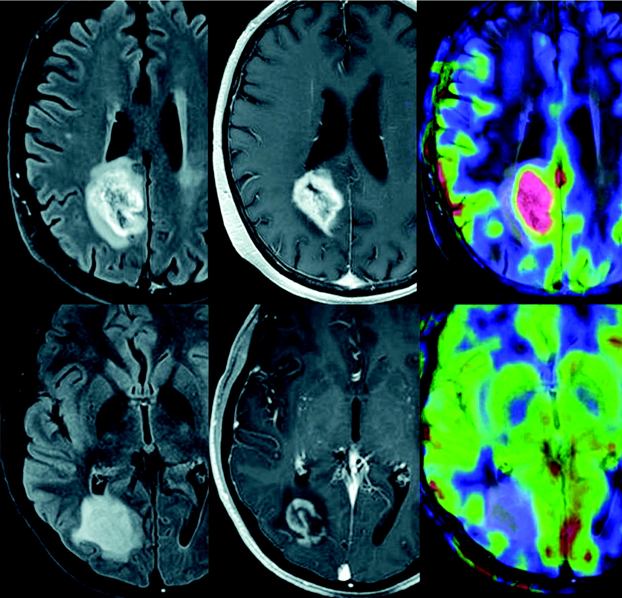
From a “visual design” perspective, MRI excels in depicting the “soft tissues” of the body, which include the brain, spinal cord, nerves, muscles, ligaments, and tendons. These structures often appear indistinct or are entirely invisible on conventional X-rays, which are primarily used for bony structures. Even CT scans, while providing good detail of bone and certain soft tissue structures, generally do not offer the same level of contrast and detail for soft tissues as MRI. For instance, MRI can exquisitely differentiate between white matter and grey matter in the brain, a level of detail unmatched by other modalities. This capability makes it invaluable for diagnosing conditions like multiple sclerosis, brain tumors, or aneurysms, where subtle changes in soft tissue are critical indicators. For injuries to the knee or shoulder, involving complex arrangements of ligaments and tendons, MRI provides definitive “high-resolution photography” that guides treatment.
Consider the role of MRI in “digital photography” of internal landscapes. While a CT scan might offer a clear “stock photo” of a general area, an MRI delivers “high-resolution” “beautiful photography” that captures the intricate textures and variations within soft tissues. This superior tissue contrast allows for the detection of subtle abnormalities that other methods might miss, making MRI an unparalleled “image tool” for comprehensive and detailed “visual design” of the body’s interior.
The Diagnostic Canvas: What MRI Reveals in Detail
The versatility of MRI is truly remarkable, allowing medical professionals to create detailed “thematic collections” of images across virtually every part of the human body. Each scan, like a unique piece of “digital art” or “beautiful photography,” offers insights critical for diagnosis and management of a vast array of conditions.
Mapping the Body: A Comprehensive Guide to MRI Applications
MRI scanners are particularly adept at providing clarity where other imaging modalities fall short, offering a comprehensive “visual design” of various bodily systems.
-
Brain and Neurological Conditions: Brain MRIs are perhaps one of the most widely recognized applications, providing “high-resolution images” of the brain’s complex structures. Neurologists rely on these detailed visuals to diagnose:
- Brain Tumors: MRI can precisely locate, measure, and characterize both cancerous and non-cancerous growths, essential for surgical planning and radiation therapy.
- Stroke: Early detection of changes in brain tissue due to stroke is crucial, and MRI’s sensitivity allows for timely intervention.
- Multiple Sclerosis (MS): Lesions—damaged areas in the brain and spinal cord—characteristic of MS are clearly visible on MRI scans.
- Aneurysms: These dangerous bulges in blood vessels can be identified before they rupture, allowing for preventative treatment.
- Epilepsy: MRI can uncover structural brain abnormalities that may be the underlying cause of seizures.
- Functional MRI (fMRI): A specialized form of MRI, fMRI observes which areas of the brain “activate” (consume more oxygen) during various cognitive tasks. This is like capturing “trending styles” of brain activity, offering “image inspiration” for understanding brain organization and assessing neurosurgical risks.
-
Spinal and Musculoskeletal Issues: For patients experiencing chronic back pain or joint problems, MRI provides invaluable “aesthetic” clarity of the spine and surrounding soft tissues. It’s the go-to “photography” method for:
- Herniated Discs: MRI clearly shows when a disc has slipped, compressing nerves and causing pain.
- Arthritis: Detailed joint images help diagnose types and severity of arthritis.
- Torn Ligaments, Tendons, and Muscles: Common in sports injuries, these soft tissue damages are expertly visualized by MRI, guiding repair strategies.
- Bone Infections (Osteomyelitis): MRI can detect infections within bones and adjacent soft tissues, particularly useful in areas like the spine.
-
Heart and Blood Vessel Conditions: Cardiac MRI offers a unique “digital art” perspective into the heart’s structure and function, helping diagnose and monitor:
- Heart Disease: Conditions like cardiomyopathy or coronary artery disease are evaluated through detailed images of heart chambers, valves, and blood flow.
- Congenital Heart Defects: Structural problems present from birth can be precisely mapped.
- Atherosclerosis: Assessment of blood flow and plaque buildup in blood vessels.
- Aneurysms: Detection of abnormal bulges in blood vessels throughout the body.
-
Abdominal and Pelvic Conditions: When ultrasound or CT scans offer insufficient information, abdominal and pelvic MRIs provide critical “high-resolution” details for:
- Liver Disease: Cirrhosis, fatty liver, and liver cancer are clearly visualized.
- Kidney Issues: Detection of kidney stones, tumors, and other abnormalities.
- Gallbladder Problems: Identification of gallstones or inflammation.
- Reproductive Health Issues: Pelvic MRI diagnoses uterine fibroids, endometriosis, ovarian cysts in women, and prostate issues in men, offering clear “backgrounds” for these delicate organs.
-
Cancer Detection and Staging: MRI plays a pivotal role in the comprehensive “image inspiration” for cancer diagnosis and management. Its ability to provide detailed 3D images helps doctors determine:
- Tumor Location and Size: Tumors in almost any part of the body can be detected and precisely measured.
- Spread (Staging): MRI helps determine how far cancer has spread, essential for accurate staging and treatment planning.
- Treatment Monitoring: Post-treatment scans check for tumor response, whether it has shrunk, grown, or spread, offering a vital “visual design” for ongoing patient care. Given its non-invasive nature, MRI is a safer option for repeated “photography” needed for cancer patients.
Beyond Diagnosis: Monitoring and Visualizing Treatment Progress
The utility of MRI extends far beyond initial diagnosis, serving as a dynamic “image tool” for tracking health journeys and inspiring “creative ideas” for patient management. The ability to perform repeated scans without radiation exposure makes it an ideal instrument for longitudinal studies and ongoing patient care.
Consider the application of MRI in monitoring chronic conditions. For patients with liver disease, the evolution of a groundbreaking NIBIB-funded project called Magnetic Resonance Elastography (MRE) represents a new frontier in “visual design” for diagnosis. This technique employs sound waves, pulsed through the liver, which the MRI then detects to determine tissue density and health. It essentially converts sound into a highly detailed “image” of tissue stiffness, capable of recognizing minute differences indicative of tumors or cirrhosis. This non-invasive method offers a safer, more comfortable, and often less expensive alternative to traditional biopsies, embodying a “creative idea” for medical imaging.
Another area of significant “image inspiration” is improving MRI accessibility and quality for challenging patient groups, particularly children. Obtaining clear MRI images in pediatric patients is notoriously difficult due to motion. Researchers are developing robust pediatric MRI systems with specialized coils designed for smaller bodies. These innovations aim to produce clearer, faster images with less operator skill, making MRIs cheaper, safer, and more available for children. Furthermore, advanced motion correction systems are being developed, acting like “AI upscalers” for real-time image stabilization, adapting MRI pulses to a patient’s movements. This “optimizer” reduces the need for repeat scans and anesthesia, benefiting both children and adults who struggle to remain still, enhancing the overall “digital photography” experience.
Beyond structural visualization, NIBIB-funded research is pushing MRI’s capabilities into the metabolic realm. While traditional MRI images structure, new techniques can measure tumor aggressiveness. By injecting specialized compounds (hyperpolarized carbon 13) into prostate cancer patients, researchers can measure the metabolic rate of a tumor. This provides a fast, accurate “photography” of its biological activity, moving beyond mere size to understand its potential for growth and spread. This information is crucial for guiding treatment decisions, especially for patients adopting a “wait and watch” approach, providing them with essential “mood boards” of their disease progression. Such innovations illustrate how MRI is not just about static “images” but about capturing dynamic, functional “trending styles” of human health at a molecular level.
Navigating the Imaging Journey: Considerations for Your Scan
Just as a photographer prepares their equipment and environment for an optimal shoot, there are several important considerations and preparations for an MRI scan to ensure the best possible “images” and a comfortable experience. Understanding these aspects helps patients approach their scan with confidence, much like a meticulous “visual designer” ensures every element of a project is perfect.
Safety and Comfort: Ensuring a Positive Imaging Experience
While MRI is celebrated for its safety due to the absence of ionizing radiation, it utilizes an extremely powerful magnetic field that necessitates specific precautions. This magnetic field, which extends beyond the machine itself, exerts strong forces on ferromagnetic objects. Therefore, patients must inform their healthcare providers about any medical implants or metal objects in their body prior to an MRI. These include pacemakers, certain neurostimulators, cochlear implants, and even some metallic joint prostheses, as the magnetic field can affect their function or cause displacement. This careful screening acts like a “filter” in “photo manipulation,” ensuring only safe elements are present to achieve a clear, unobstructed “image.”
The MRI environment can also present sensory challenges. The machine often produces loud clicking and beeping sounds, sometimes reaching up to 120 decibels. Patients are always provided with earplugs or headphones, often with music, to protect their hearing and help manage the noise—creating a more pleasant “aesthetic background” for the procedure. Some patients may also experience a twitching sensation from the rapidly switched magnetic fields, which, while harmless, can be startling.
For enhanced “image” quality, contrast agents may be used. While generally safe, there’s a rare risk of allergic reactions, usually mild. More significantly, patients with severe renal failure who require dialysis face a very rare but serious condition called nephrogenic systemic fibrosis (NSF) linked to certain gadolinium-containing agents. Current guidelines recommend cautious use of contrast in such cases, with dialysis performed promptly post-scan to remove the agent. This highlights that even the “editing styles” of medical imaging require careful consideration.
Pregnancy is another consideration. While no definitive harmful effects on the fetus have been demonstrated, MRI scans are generally avoided as a precaution, especially during the first trimester when organs are forming, and if contrast agents are considered, due to potential fetal exposure. This adheres to a principle of “visual design” caution, ensuring minimal theoretical risk.
Claustrophobia is a common concern, as traditional MRI machines are enclosed tunnels. To mitigate this, many facilities offer “open MRI” scanners, which are open on the sides rather than fully enclosing the patient. This represents a significant “visual design” adaptation for patient comfort, especially for those who find narrow spaces distressing or whose size makes traditional machines impractical. For other patients, strategies like visualization techniques, sedation, or even anesthesia are available, transforming a potentially anxiety-inducing experience into one that is calm and manageable, allowing them to relax and focus on creating their “inner wallpaper.”
Preparing for Your MRI: A Guide to Optimal Imaging
Effective preparation is key to obtaining “high-resolution” and diagnostically valuable MRI “images.” Much like setting up for a professional “photography” shoot, certain steps ensure the best results.
Before the scan, patients will typically be asked to change into a hospital gown or scrubs. This is to eliminate any clothing items that might contain metal, such as zippers, buttons, or even certain performance fabrics with metallic threads that can heat up in the magnetic field. All jewelry, watches, piercings, removable dental work, and other metallic accessories must be removed. These objects can interfere with the magnetic field, distort the “image,” or become dangerous projectiles. This meticulous removal process ensures a “clean canvas” for the internal “digital photography.”
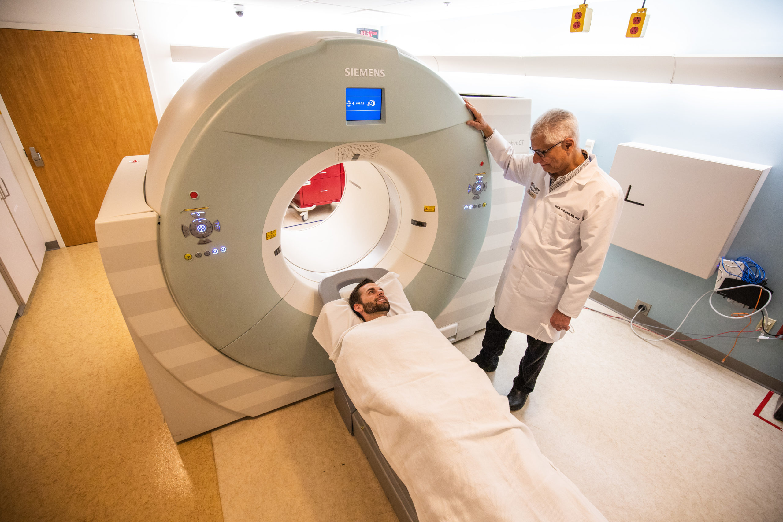
Patients will lie on a scanning bed that slides into the MRI machine. The importance of remaining absolutely still during the entire procedure, which can range from 20 to 60 minutes depending on the area being scanned, cannot be overstated. Movement can cause “blurring” in the “images,” requiring repeat sequences and extending scan time. Technologists closely monitor patients throughout the scan, and an intercom system allows for two-way communication, providing reassurance and allowing patients to voice any discomfort.
The MRI technologist, a healthcare provider specially trained to operate the MRI scanner, works in conjunction with a radiologist, a medical doctor who interprets the “images” and compiles a detailed report. This report, essentially an “Image-to-Text” conversion, is then sent to the referring healthcare practitioner, typically within 24-48 hours. This swift delivery ensures that critical “visual design” insights are quickly translated into actionable medical decisions.
The process of booking an MRI, typically requiring a referral from a physician, varies by location and healthcare system. In some regions, wait times for publicly funded MRIs can be extensive, while private clinics may offer faster appointments, albeit at a cost. For example, at Tophinhanhdep.com, we acknowledge that the desire for clear “images” extends to all aspects of life, including health. Understanding these practicalities, from referral requirements to potential costs, is part of navigating the journey towards comprehensive internal “visual design” and better health management.
In conclusion, Magnetic Resonance Imaging is a marvel of modern medicine, an advanced “image tool” that provides an unparalleled window into the human body. From its fundamental physics, converting subtle proton signals into “high-resolution digital photography,” to its diverse applications in diagnosing and monitoring a vast array of conditions, MRI continuously offers new “image inspiration” for healthcare professionals. Just as Tophinhanhdep.com strives to deliver exquisite “images,” “photography,” and “visual design” resources, MRI delivers clarity and detail vital for understanding our most intricate internal “landscapes,” empowering us to safeguard our health with precision and insight.