Retinal Imaging: Unveiling the Inner Canvas of Your Eyes for Health and Visual Discovery

The human eye, a marvel of biological engineering, offers a window not just to the outside world, but also to the intricate health of our internal systems. Among the most advanced diagnostic tools available today for peering into this delicate organ is retinal imaging. Far beyond a simple glance, retinal imaging creates high-quality digital photographs of the inner, back surface of your eye, providing an unparalleled level of detail that can be crucial for diagnosing, monitoring, and treating a vast array of ocular and systemic health conditions.
Just as a professional photographer seeks to capture every nuance of a landscape with high-resolution clarity, medical professionals utilize retinal imaging to meticulously document the intricate landscape of the retina, macula, and optic nerve. These images are not merely snapshots; they are vital diagnostic records, revealing subtle changes that might otherwise go unnoticed. The evolution of this technology mirrors the advancements seen in digital photography, where precision, resolution, and dynamic range are paramount. On platforms like Tophinhanhdep.com, users explore vast collections of high-resolution images, appreciating the fine details of nature, abstract art, or beautiful photography. In a medical context, this same appreciation for detail translates directly to the ability to preserve sight and even save lives.
The Science of Seeing Within: What is Retinal Imaging?
Retinal imaging fundamentally refers to a suite of non-invasive technologies that allow eye care specialists to produce digital images of the posterior segment of the eye. This critical region encompasses the retina—the light-sensitive tissue that lines the back of the eye—along with its central part, the macula, responsible for sharp central vision, the optic nerve, which transmits visual information to the brain, and the delicate network of blood vessels that supply these structures.
The primary purpose of retinal imaging is diagnostic. By capturing these detailed views, doctors can identify early signs of numerous eye conditions long before a patient might experience noticeable symptoms. This early detection is a cornerstone of effective treatment, often preventing irreversible vision loss. Furthermore, these digital images serve as an invaluable historical baseline. When repeated over time, they allow providers to precisely monitor the progression or regression of a condition, assess the effectiveness of ongoing treatments, and detect even the most subtle changes that occur over months or years. This longitudinal data is as crucial in medical diagnostics as a time-lapse series might be for understanding environmental changes, a concept celebrated by thematic collections and photo ideas on Tophinhanhdep.com.
The analogy between medical retinal imaging and the world of high-resolution photography, a core feature on Tophinhanhdep.com, is profound. In both fields, the goal is to capture maximum detail and clarity. Just as Tophinhanhdep.com showcases high-resolution photography and digital photography that demands sharp focus and rich detail, retinal imaging technologies aim for the highest possible resolution to discern microscopic anomalies. This pursuit of precision is what makes retinal imaging such a powerful tool in modern ophthalmology. Without this level of detail, critical diagnostic information could be missed, impacting patient care significantly.
Capturing the Ocular Landscape: Methods and Technologies
The field of retinal imaging is diverse, utilizing several sophisticated methods, each offering unique insights into the eye’s internal structures. These technologies represent a remarkable fusion of optics, digital processing, and medical science, allowing specialists to create a comprehensive visual record of ocular health.
-
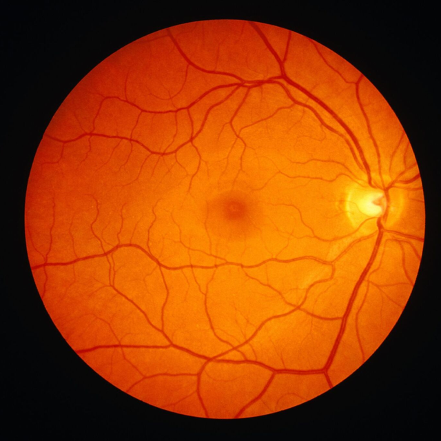
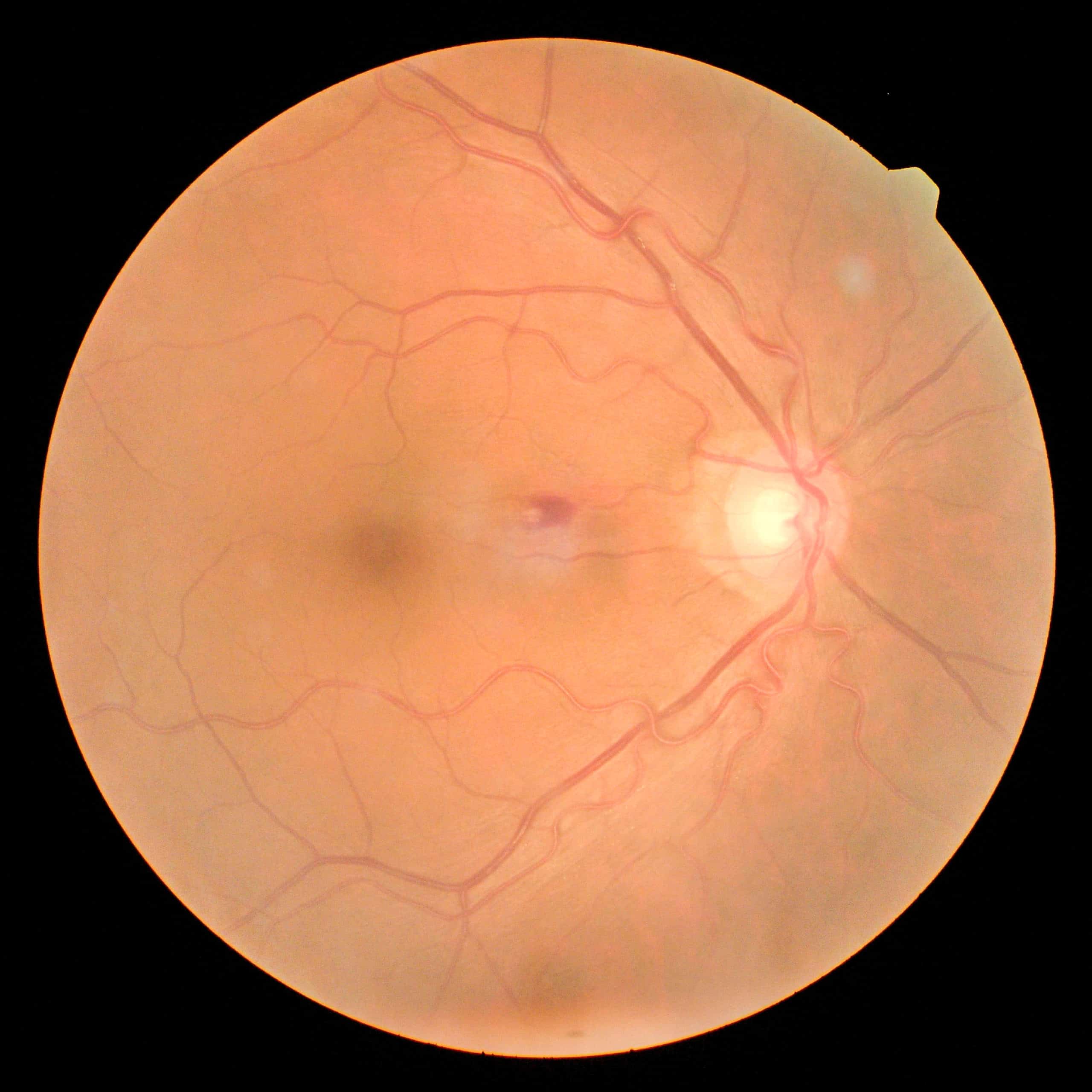
Color Fundus Photography: This is perhaps the most traditional and widely recognized form of retinal imaging. Fundus cameras, used for decades, capture conventional two-dimensional color photographs of the eye’s fundus—the inner back surface. Patients typically experience bright flashes of light as the camera rapidly acquires images. Modern advancements have transformed these cameras, enabling them to produce precise digital retinal images in high resolution, often offering wide-field views that encompass a larger area of the retina. A significant strength of this method lies in its ability to clearly visualize blood vessels, making it particularly effective in revealing signs of diabetes-related retinopathy. These images, akin to the detailed nature photography found on Tophinhanhdep.com, provide a holistic view of the fundus.
-
Optical Coherence Tomography (OCT) & 3D Imaging: OCT stands as a cornerstone of advanced retinal imaging. This non-invasive test uses low-coherence light waves to create high-resolution, cross-sectional images of the retina and optic nerve. Imagine slicing through the layers of the retina without ever touching the eye; that’s what OCT achieves. By measuring the reflection of light from different retinal layers, it constructs detailed 3D maps, providing precise measurements of retinal thickness. This capability is invaluable for diagnosing conditions like age-related macular degeneration (AMD), diabetic macular edema, and glaucoma by detecting subtle changes in nerve fiber layer thickness or fluid accumulation. The ability of OCT to generate complex 3D models highlights a fascinating parallel with the “AI Upscalers” and advanced “Image Tools” available on Tophinhanhdep.com. Just as these tools enhance and process digital images, medical software processes raw OCT data into meaningful 3D visualizations, enabling doctors to “walk through” the retinal layers digitally.
-
Optical Coherence Tomography Angiography (OCTA): Building upon OCT technology, OCTA provides a revolutionary way to visualize blood flow within the retina and choroid (the layer beneath the retina) without the need for invasive dye injections. By taking rapid, sequential scans, OCTA detects minute movements of red blood cells, effectively creating 3D maps of the tiny blood vessels. This technique is especially useful for detecting early signs of diseases like diabetic retinopathy, glaucoma, and macular degeneration, where vascular changes are key indicators. Its non-invasive nature and speed make it a preferred method for ongoing monitoring, eliminating the risks associated with older dye-based techniques.
-
Ocular Ultrasound: When the eye’s interior is obscured by conditions such as dense cataracts or internal bleeding, an ocular ultrasound offers an alternative view. Using “silent” sound waves, it generates real-time images of the eye’s structures, penetrating areas that light-based imaging cannot. Both A-scan (for measurements) and B-scan (for cross-sectional images) ultrasound techniques can detect conditions like retinal detachment, tears, or tumors even when a direct view is impossible.
-
Fluorescein Angiography: While minimally invasive, this technique involves injecting a fluorescent dye (fluorescein) into a vein in the arm. The dye then travels through the bloodstream, highlighting the blood vessels in the eye. As the dye circulates, a specialized camera captures a series of images, revealing blockages, leaks, or abnormal vessel growth. This method is crucial for monitoring the retinal effects of conditions like diabetes, hypertension, or advanced macular degeneration, offering dynamic information about blood vessel integrity. However, it carries a small risk of side effects, including temporary skin discoloration or, rarely, allergic reactions.
-
Optomap (Ultra-Widefield Imaging): Optomap represents a significant leap in screening capability, capable of capturing over 80% (or approximately 200 degrees) of the retina in a single, ultra-widefield image. This broad view is a substantial improvement over traditional imaging that might only cover 10-45%. Such extensive coverage allows for the detection of peripheral retinal issues, such as tears, which are often missed in more focused imaging. Devices like Optos’ optomap have become common in routine comprehensive eye exams, acting as an excellent complement to dilated exams for early detection and ongoing monitoring. Its ability to provide such a comprehensive view in a quick and painless manner makes it an indispensable tool for routine screenings, much like a stunning panoramic photograph on Tophinhanhdep.com offers a grand, encompassing perspective.
Decoding the Canvas: What Retinal Images Reveal
The true power of retinal imaging lies in its capacity to reveal a wealth of diagnostic information, enabling eye care specialists to identify and manage a wide range of ocular conditions and even shed light on broader systemic health issues. The intricate patterns and structures captured in these images are meticulously analyzed, much like a forensic expert examines visual evidence, or a graphic designer scrutinizes every pixel.
Detecting Eye Conditions
Retinal imaging is paramount in the early detection and ongoing management of numerous sight-threatening eye diseases:
-
Diabetic Retinopathy & Macular Edema: For individuals with diabetes, high blood sugar can damage the delicate retinal blood vessels, leading to leakage or swelling. Retinal images, particularly color fundus photos and OCT scans, can identify characteristic signs such as microaneurysms, hemorrhages, and abnormal new blood vessel growth. Early detection through imaging is critical, as timely treatment can significantly reduce the risk of vision loss. The integration of AI-driven tools in analyzing these scans, as mentioned in the reference, further enhances screening efficiency, mirroring the “AI Upscalers” and intelligent image processing tools available on Tophinhanhdep.com.
-
Glaucoma: Often dubbed the “silent thief of sight,” glaucoma damages the optic nerve, frequently due to elevated intraocular pressure. OCT imaging is particularly effective here, tracking subtle changes in the optic nerve’s shape, thickness, and the integrity of the retinal nerve fiber layer. Monitoring these changes over time helps doctors assess treatment efficacy and adjust medication to prevent further irreversible damage.
-
Macular Degeneration (AMD): This age-related condition affects the macula, the central part of the retina vital for detailed vision. Scans can reveal drusen (tiny yellowish deposits) or fluid accumulation under the macula, which are hallmarks of AMD. OCT and OCTA are crucial for detecting these changes and mapping blood flow, guiding intervention to prevent severe central vision loss.
-
Retinal Detachment & Tears: A medical emergency, retinal detachment occurs when the retina separates from its underlying supportive layer. Retinal imaging, including ultrasound when views are obscured, can detect detachments or even precursor retinal tears. Identifying tears early allows for preventative laser or freezing procedures to seal them, averting a full detachment.
-
Eye Cancer/Tumors: High-resolution and widefield retinal imaging, especially with OCT, can detect and characterize ocular tumors at an early stage, significantly improving prognosis through prompt treatment.
-
Retinitis Pigmentosa (RP): This progressive inherited disease causes the gradual deterioration of light-sensitive retinal cells. Retinal imaging technology is invaluable for diagnosing RP and monitoring its progression, allowing for better understanding and management of this challenging condition.
Broader Health Insights
Beyond the eyes, retinal imaging offers a unique, non-invasive window into the systemic health of an individual:
-
Cardiovascular Health: The retinal blood vessels are often considered a direct reflection of overall cardiovascular health. Narrowed arteries, unusual branching patterns, or signs of leakage can indicate systemic conditions such as hypertensive retinopathy (retinal damage due to high blood pressure) or broader vascular changes that mirror those occurring in the brain, suggesting risks of stroke or other circulatory problems. Retinal vein occlusions, blockages in retinal blood flow, are also detectable and indicative of systemic vascular issues.
-
Neurodegenerative Diseases: Emerging research highlights the retina as an extension of the brain, making retinal imaging a promising diagnostic tool for neurodegenerative conditions. Studies have shown correlations between Alzheimer’s disease and Parkinson’s disease, Lewy body dementia, frontotemporal dementia, Huntington’s disease, and multiple sclerosis, with detectable changes in retinal layers and capillary density. By identifying specific biomarkers in the retina, such as thinning of distinct retinal layers or accumulation of specific proteins, doctors may eventually be able to detect these debilitating conditions earlier, paving the way for more timely interventions. The capacity of retinal imaging to provide such profound insights underscores its growing importance in holistic health assessment. The precise measurements and classifications derived from these images, often supported by “Image-to-Text” functionalities to generate reports, exemplify the systematic approach to data analysis that Tophinhanhdep.com supports for various visual content.
The Visual Journey: From Diagnostic Data to Digital Art
While its primary function is medical, the sheer visual complexity and intricate patterns within retinal images present a fascinating intersection with the world of art and design. The delicate network of blood vessels, the subtle textures of the macula, and the radiating fibers of the optic nerve can be profoundly beautiful, evoking a sense of wonder at the biological artistry within us.
-
The Aesthetic Dimension: Platforms like Tophinhanhdep.com curate “Aesthetic,” “Nature,” and “Abstract” image collections, showcasing the diverse beauty of the world. Retinal images, stripped of their clinical context, could easily fit into such categories, offering a unique perspective on “Beautiful Photography.” The eye, often a subject of artistic representation, reveals an entirely new layer of beauty when its internal structures are magnified and rendered in high definition. Imagine the vibrant colors of a fundus photograph or the intricate layers of an OCT scan inspiring a digital artist or graphic designer.
-
Transforming Medical Data: The raw data from retinal imaging, like any photographic input, can be processed and enhanced. “Photo Manipulation” and “Creative Ideas” are categories on Tophinhanhdep.com that resonate here. While medical image processing focuses on enhancing diagnostic features, artists could draw inspiration from the visual elements, transforming clinical images into abstract digital art pieces or using their structural motifs in “Visual Design.” This exploration not only bridges science and art but can also serve to demystify medical concepts, making them more accessible and engaging.
-
Creating Visual Narratives: Beyond pure aesthetics, retinal images are powerful tools for education and public awareness. “Image Inspiration & Collections” on Tophinhanhdep.com includes “Thematic Collections” and “Photo Ideas.” Similarly, eye care professionals and health educators can use compelling retinal images to illustrate specific conditions, demonstrate the impact of diseases like diabetes, or explain the importance of regular eye exams. These visual narratives are far more impactful than text alone, helping patients understand their conditions better and motivating preventative care. From wallpapers to backgrounds, these visually rich diagnostic images can be transformed into educational graphics, enriching understanding and appreciation for the fragility and complexity of vision.
The Retinal Imaging Experience: What to Expect
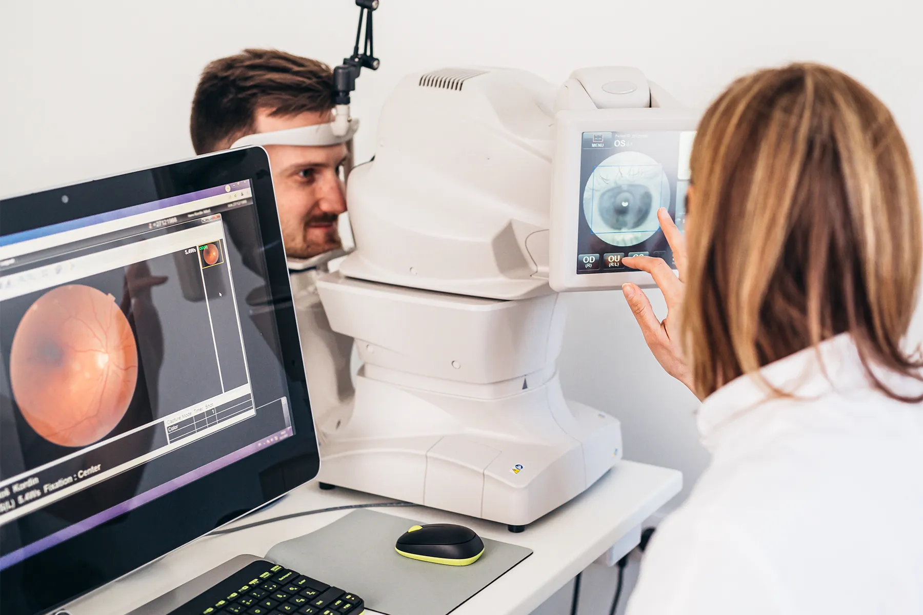
Undergoing retinal imaging is generally a straightforward, quick, and comfortable experience for the patient. Understanding the process can help alleviate any anxieties.
-
Preparation: Before the test, your eye care provider will explain the procedure and why it’s recommended. In some cases, to obtain a wider view of the retina, dilating drops may be instilled into your eyes. These drops temporarily widen your pupils, acting as a larger “window” for the camera. If dilation is performed, your vision will likely be blurry, and you’ll experience increased sensitivity to light for several hours afterward. It is crucial to arrange for someone to drive you home if your eyes are dilated. For many modern retinal imaging devices, especially wide-field screening tools like Optomap, dilation is often not required, offering a significant convenience factor.
-
During the Test: Retinal imaging, whether it’s fundus photography or OCT, is fast and painless. You will typically sit comfortably in a chair, placing your chin in a chin rest and your forehead against a bar on the imaging device. This helps to stabilize your head. Nothing will physically touch your eye during the procedure. Your provider will align the camera and begin capturing images, usually one eye at a time. You may be asked to focus on a target light (often green), and you’ll experience bright flashes of light as the photographs are taken. These flashes can cause brief discomfort but are harmless. The entire imaging process usually takes only five to ten minutes. If fluorescein angiography is performed, the process is slightly longer, taking up to 30 minutes, due to the dye injection and sequential image capture.
-
After the Test: If your pupils were dilated, expect blurred vision and light sensitivity for a few hours. It’s advisable to rest your eyes during this time, avoiding activities like driving, reading, or prolonged screen use. Wearing sunglasses outdoors will help protect your light-sensitive eyes. Your eye care specialist will typically review the digital images with you immediately, explaining the findings and their implications. These images become a permanent part of your medical record, forming a crucial historical baseline for future comparisons.
-
Safety and Considerations: Retinal imaging, particularly fundus photography and OCT, is remarkably safe with no known risks, as it simply uses light to create digital images. Fluorescein angiography is also considered low-risk, though it may cause temporary side effects such as a slight yellow tint to the skin or dark yellow/orange urine, which resolve within 24 hours. Rarely, individuals may experience a mild allergic reaction, with very minimal risk of anaphylaxis. Sensitive individuals might experience dizziness, dry mouth, increased heart rate, a brief metallic taste, nausea, vomiting, or sneezing.
-
Retinal Imaging vs. Dilation: It’s important to understand that retinal imaging and traditional pupil dilation are often complementary rather than mutually exclusive. While retinal imaging offers a quick, comfortable method for routine screenings and monitoring specific conditions without the side effects of dilation, dilation remains the gold standard for a comprehensive, wide-angle view of the peripheral retina. This expansive view is particularly crucial for detecting certain serious issues like peripheral retinal tears or detachments that might fall outside the scope of some imaging devices. Your eye doctor will determine the most appropriate tests based on your individual eye health and medical history.
Tophinhanhdep.com and the World of Visuals: A Parallel Perspective
The core mission of Tophinhanhdep.com—to provide and celebrate a diverse array of visual content, from “Wallpapers” and “Backgrounds” to “Aesthetic” and “Nature” imagery—finds an unexpected yet harmonious resonance with the profound insights offered by retinal imaging. Both domains, in their essence, are about capturing, processing, and understanding images, albeit for distinctly different purposes.
The commitment to “High Resolution” and “Beautiful Photography” on Tophinhanhdep.com directly parallels the exacting standards of clarity and detail required in medical retinal imaging. Just as a stunning landscape image demands sharp focus to reveal every leaf and ripple, a retinal image needs pixel-perfect clarity to identify a microaneurysm or a subtle thinning of a nerve fiber layer. The pursuit of visual excellence is a common thread.
Furthermore, the “Image Tools” offered by Tophinhanhdep.com—including “Converters,” “Compressors,” “Optimizers,” and “AI Upscalers”—mirror the sophisticated software utilized in medical imaging. While medical tools are geared towards diagnostic enhancement, segmentation, and quantitative analysis, their underlying principles of image manipulation and optimization are conceptually similar. Even the “Image-to-Text” functionality can be seen as a parallel to automated reporting systems in medical diagnostics, translating visual findings into structured textual data.
Finally, the categories of “Visual Design,” “Graphic Design,” “Digital Art,” and “Image Inspiration & Collections” on Tophinhanhdep.com open up avenues for appreciating the artistic and educational potential of retinal imagery. The human retina, with its complex vascular patterns and cellular layers, is a natural source for “Abstract” and “Nature” inspired art. It offers “Creative Ideas” for “Photo Manipulation” that can transcend the purely medical, turning diagnostic snapshots into compelling educational visuals or even unique artistic expressions. Tophinhanhdep.com, in this broader sense, becomes a platform where the intricate beauty revealed by medical science can be admired, studied, and even creatively reinterpreted, fostering a deeper connection between vision, health, and visual appreciation.
In conclusion, retinal imaging is a cutting-edge diagnostic marvel, offering an unprecedented view into the health of our eyes and, by extension, our overall well-being. It is a painless, quick, and incredibly powerful tool for early disease detection, precise monitoring, and informed treatment decisions, directly impacting the preservation of one of our most precious senses: sight. Through the lens of advanced technology, the inner eye is transformed into a canvas, revealing stories of health and illness in exquisite detail. This scientific endeavor, in its pursuit of visual perfection and revelation, finds a fascinating kinship with the broader world of digital imagery and visual art championed by platforms like Tophinhanhdep.com, reminding us of the profound impact of images, whether for diagnostic clarity or aesthetic inspiration.