What is SPECT Imaging? A Journey into the Art and Science of Inner Visuals

In an era dominated by digital imagery, from breathtaking wallpapers to intricate graphic designs, the pursuit of capturing and interpreting visual information has reached unprecedented levels. While our daily lives are saturated with images, there exists a profound realm of visuals that transcends ordinary perception: the internal landscape of the human body. Among the most fascinating techniques for revealing this hidden world is Single-Photon Emission Computed Tomography, widely known as SPECT imaging. Far from being a mere diagnostic tool, SPECT imaging offers a unique form of “inner photography,” generating 3D images that provide critical insights into physiological function. For enthusiasts of high-resolution imagery, digital art, and visual design, understanding SPECT imaging not only offers a glimpse into advanced medical science but also provides a novel source of abstract, functional aesthetics.
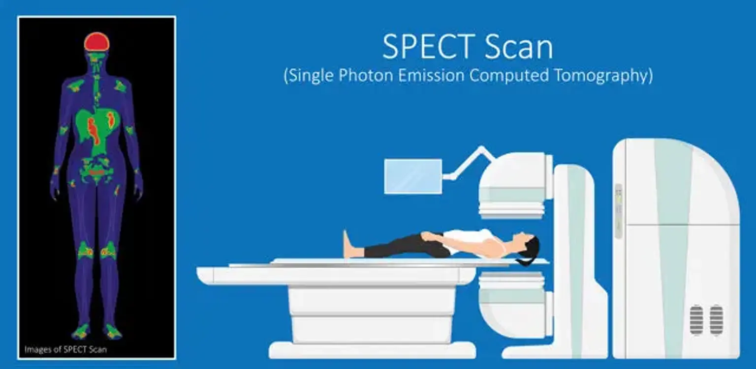
At its core, SPECT is a non-invasive nuclear imaging test that allows medical professionals to visualize how internal organs are functioning. Unlike traditional X-rays, which offer a static picture of anatomical structures, SPECT provides dynamic insights into blood flow, metabolic activity, and cellular uptake. This capability transforms raw data into vivid, multi-dimensional pictures, akin to highly specialized digital photography of the body’s dynamic processes. Imagine capturing the subtle currents of blood flowing through the heart, or mapping the neural activity within the brain – these are the powerful visuals that SPECT brings to light.
The technology behind SPECT involves injecting a small amount of a radioactive substance, known as a radiotracer, into the patient’s bloodstream. This tracer is specifically designed to target certain molecules or biological processes within the body. As the tracer travels and accumulates in the organs of interest, it emits gamma rays. A specialized device, aptly named a gamma camera, then detects these emissions. This camera rotates around the patient, capturing a multitude of 2D projections from various angles. A sophisticated computer algorithm then processes these projections, reconstructing them into a comprehensive 3D image. This reconstruction process is a marvel of computational visual design, transforming discrete data points into an understandable and visually rich representation of internal biological activity.
For those interested in the broader context of visual information, these SPECT images, with their varying shades of color indicating different levels of tracer uptake, offer a distinctive aesthetic. They are, in a sense, abstract artworks generated by biological processes, providing a new dimension for visual inspiration and thematic collections on platforms like Tophinhanhdep.com. Just as photographers capture nature’s intricate patterns or designers craft compelling graphics, SPECT illuminates the inherent design within human physiology, opening avenues for creative interpretation and understanding.
The Science Behind the Image: How SPECT Works
The creation of a SPECT image is a meticulously orchestrated process that combines nuclear medicine, advanced camera technology, and sophisticated computational reconstruction. Understanding these foundational elements not only demystifies the scan but also highlights the complexity involved in generating such unique visual data. It’s a testament to digital imaging capabilities, providing a window into the otherwise unseen.
Tracers and Gamma Cameras: The Heart of Visual Data Capture
The journey of a SPECT image begins with the radiotracer. These are not merely generic radioactive materials but carefully selected substances tailored to bind to specific molecules or accumulate in particular tissues. For instance, in cardiac imaging, tracers are chosen that are absorbed by healthy heart muscle, allowing physicians to assess blood flow. Once injected intravenously, the tracer mixes with the patient’s blood and begins its circulation, becoming “taken up” or absorbed by the living tissues.
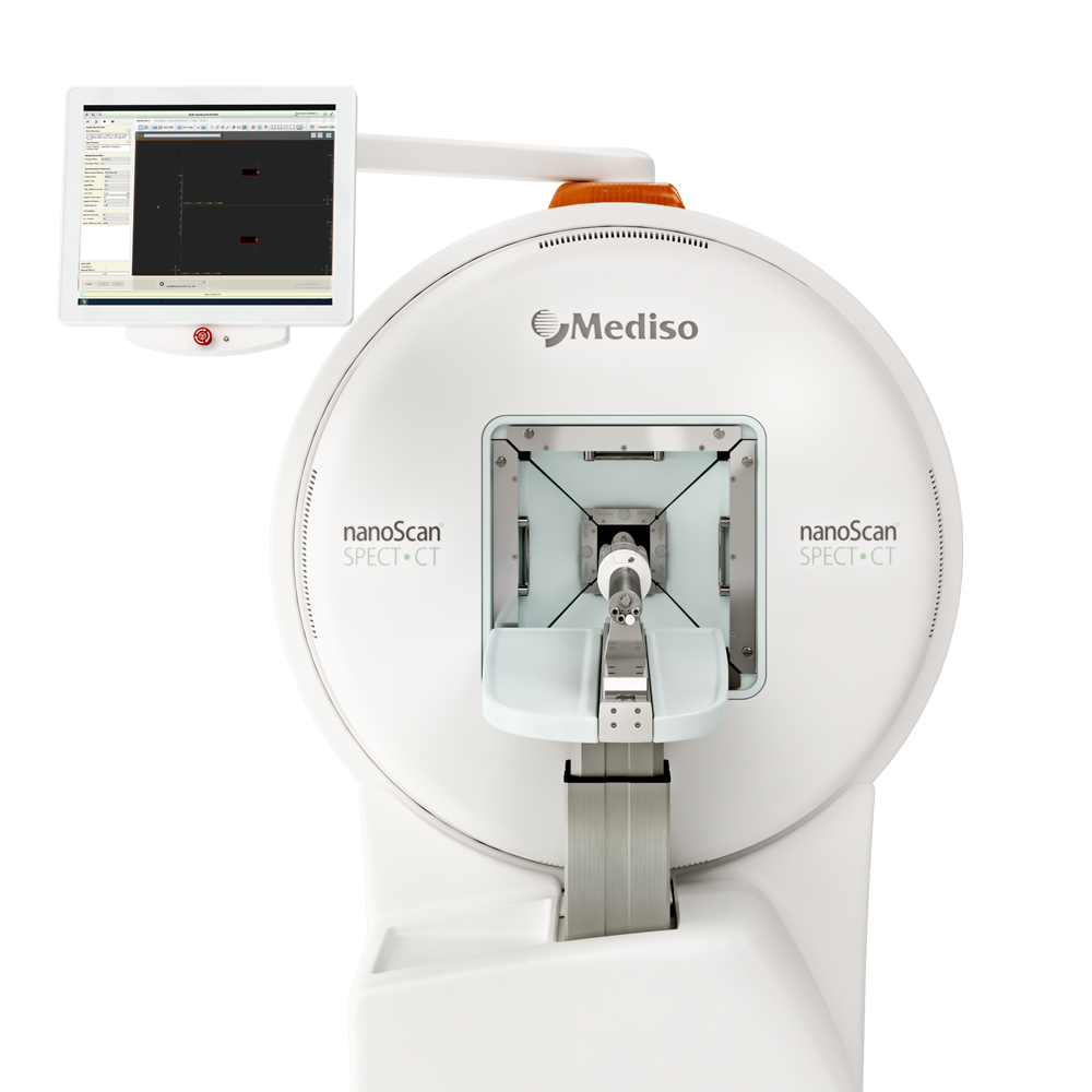
As these tracers decay, they emit single photons of gamma radiation. This is where the specialized gamma camera comes into play. Modern gamma cameras, such as those employing advanced solid-state technology with silicon-based photodiodes and cesium iodide (CsI), offer superior sensitivity and high energy resolution. This technological advancement means not only clearer image acquisition but also allows for smaller, more patient-friendly designs compared to older systems or larger MRI machines. These cameras don’t just take a single picture; they continuously detect the gamma rays as they rotate around the patient, typically acquiring projections every 3-6 degrees for a full 360-degree rotation. The time taken for each projection can vary, but usually lasts about 15-20 seconds, resulting in a total scan time of 15-20 minutes. Multi-headed cameras can expedite this process, capturing multiple projections simultaneously. This array of collected data forms the raw input for the visual reconstruction.
Reconstructing Reality: From 2D Projections to 3D Masterpieces
The sheer volume of 2D projection data collected by the gamma camera is then fed into a powerful computer. This is where the magic of image processing and visual design truly manifests. Using sophisticated algorithms, the computer “reconstructs” these numerous 2D images into a detailed 3D representation of the organ or area being examined. This process is analogous to how digital artists create 3D models from multiple photographic perspectives or how image tools on Tophinhanhdep.com might convert or optimize visual data.
The resulting 3D dataset is remarkably versatile. It can be manipulated to display thin slices along any chosen axis of the body – sagittal, coronal, or axial – much like other tomographic techniques such as MRI, CT, and PET scans. This multi-planar viewing capability allows physicians to explore the internal structures and their functional activity from every conceivable angle, providing a comprehensive visual understanding of the biological processes underway. The different shades of color appearing in these reconstructed images are not arbitrary; they directly correlate to the concentration of the radioactive tracer in various areas. Brighter regions indicate higher tracer uptake and, consequently, greater physiological activity or blood flow, while darker areas signify reduced uptake, potentially pointing to compromised function or damage. This visual language, a sophisticated form of digital art, enables medical professionals to interpret complex biological data with remarkable precision.
Unveiling Hidden Narratives: Applications of SPECT Imaging
The ability of SPECT imaging to visualize physiological function rather than just anatomical structure makes it an indispensable tool in modern medicine. Its applications span various medical specialties, each benefiting from the unique “visual narratives” these scans provide about the body’s inner workings. These images offer unique insights, much like high-resolution stock photos capture specific moments or abstract art conveys complex emotions.
Cardiac Visuals: Mapping the Heart’s Dynamic Blood Flow
One of the most common and critical applications of SPECT imaging is in cardiology, specifically for Myocardial Perfusion Imaging (MPI) or nuclear stress tests. This technique allows physicians to assess blood flow to and from the heart muscle, identifying restrictions or blockages in the coronary arteries. The process often involves taking two sets of images: one while the patient is exercising (or chemically stressed if they cannot exercise) and another while at rest.
By comparing these “stress” and “rest” images, medical professionals can visually evaluate how well the heart muscle is perfused under different levels of exertion. Areas of the heart receiving adequate blood flow will appear bright and well-defined, indicating normal function. Conversely, regions with reduced or absent blood flow will appear darker or as “cold spots,” signifying insufficient blood supply due to narrowed or clogged arteries, or even scar tissue from a previous heart attack. This side-by-side visual comparison is crucial for diagnosing coronary artery disease, evaluating the success of interventions like bypass surgery, and guiding treatment plans. The visual clarity and functional detail provided by cardiac SPECT scans are akin to high-resolution digital photography that captures a dynamic physiological event, offering a diagnostic precision that traditional X-rays cannot match.
Neurological Insights: Picturing Brain Function
Beyond the heart, SPECT imaging plays a vital role in evaluating a range of brain and neurological conditions. The brain, with its complex network of neurons and intricate blood supply, benefits significantly from SPECT’s ability to visualize functional activity. Tracers used in neurological SPECT scans are designed to cross the blood-brain barrier and accumulate in brain tissue in proportion to blood flow or receptor density.
These scans can provide crucial information for diagnosing and monitoring conditions such as:
- Alzheimer’s disease and other dementias: SPECT can show patterns of reduced blood flow in specific brain regions associated with these degenerative conditions, often before structural changes are visible on other imaging modalities like MRI. These images offer an “aesthetic” yet poignant representation of cognitive decline.
- Epilepsy: During a seizure, blood flow to the affected brain region increases. Performing a SPECT scan during or shortly after a seizure can pinpoint the exact origin of the epileptic activity, aiding in surgical planning.
- Parkinson’s disease: Specialized SPECT tracers (e.g., DaTscan) can visualize dopamine transporters in the brain, helping to differentiate Parkinson’s disease from other movement disorders.
- Stroke and traumatic brain injury: SPECT can assess blood flow to damaged brain tissue, helping to determine the extent of injury and potential for recovery.
- Psychiatric disorders: While still largely research-based, SPECT has been explored for visualizing altered blood flow patterns in conditions like depression and ADHD, offering a deeper, more abstract understanding of mental states.
The detailed 3D maps of brain activity generated by SPECT provide unique visual data that not only aid in diagnosis but also inspire a deeper appreciation for the complex interplay of biology and visual representation. For those passionate about visual design and image collections, the vibrant, often abstract patterns of brain SPECT scans could easily become part of “thematic collections” or serve as “mood boards” for creative ideas.
SPECT vs. PET: A Comparative Look at Imaging Resolution and Detail
In the realm of nuclear medicine imaging, SPECT often draws comparisons with Positron Emission Tomography (PET). Both techniques utilize radioactive tracers to produce functional images, but they differ significantly in their underlying physics, resulting in variations in image resolution, cost, and typical applications. Understanding these distinctions is crucial, much like discerning between different digital photography formats or editing styles on Tophinhanhdep.com.
Resolution and Clarity: Capturing Finer Details
The primary difference between SPECT and PET lies in the type of radioactive tracer used and how their emissions are detected. SPECT tracers emit single gamma photons directly, which are then detected by the gamma camera. While this method is highly effective, the inherent physics typically limits SPECT image resolution to approximately 10-20 millimeters. This means SPECT is excellent for visualizing larger areas of functional activity or significant anomalies but might struggle with pinpointing very small structures or subtle changes. The clarity of SPECT images can be likened to a well-composed stock photo, providing clear context and overall information.
PET scans, on the other hand, utilize tracers that emit positrons. When a positron encounters an electron in the body, they annihilate each other, producing two gamma photons that travel in precisely opposite directions. PET scanners are designed to detect these “coincident” emissions simultaneously. This coincidence detection provides much more precise localization information for the radioactive event, leading to significantly higher image resolution, typically around 5 millimeters. This enhanced resolution means PET scans are superior for detecting smaller abnormalities, such as early-stage cancer lesions or very subtle changes in brain metabolism. The detailed output of a PET scan can be compared to a high-resolution, finely-tuned digital photograph, offering minute details that might be critical for diagnosis.
Accessibility and Cost: Practical Considerations for Visual Acquisition
Beyond technical resolution, there are practical differences in the accessibility and cost of SPECT versus PET imaging. SPECT machines are generally more widely available in hospitals and diagnostic centers worldwide. This broader availability contributes to SPECT scans often being significantly less expensive than PET scans. The lower cost is partly due to the fact that SPECT can utilize longer-lived, more easily obtained radioisotopes. These isotopes can be produced off-site and transported to various facilities, reducing the need for specialized on-site infrastructure.
In contrast, PET tracers typically have very short half-lives (e.g., Fluorine-18, a common PET tracer, has a half-life of about 110 minutes), meaning they decay rapidly. This necessitates the production of PET radioisotopes in a cyclotron, often located on-site or very close to the PET scanning facility. The specialized equipment and logistical requirements for tracer production contribute to the higher operational costs and, consequently, the higher price of PET scans.
Therefore, the choice between SPECT and PET often comes down to a balance between the required image resolution for a specific diagnosis and practical considerations like cost and accessibility. For many applications, particularly in cardiology and bone imaging, SPECT provides sufficient diagnostic value at a more accessible price point. When detecting very small lesions, assessing early metabolic changes, or requiring the highest possible functional image clarity, PET’s superior resolution often makes it the preferred, albeit more expensive, option. Both technologies, however, contribute immensely to the visual data landscape of internal medicine, offering physicians and patients alike unparalleled insights into health and disease, much like diverse photographic techniques enrich our visual world.
Beyond Diagnostics: SPECT as Inspiration for Visual Creators
While the primary purpose of SPECT imaging is medical diagnosis, the unique visual outputs generated by these scans hold an untapped potential for creative expression and artistic inspiration. For a platform like Tophinhanhdep.com, dedicated to images, photography, visual design, and creative ideas, SPECT images offer a fascinating intersection of science, art, and abstract beauty. They are, in essence, a new genre of internal photography.
Abstract Beauty: Finding Art in Medical Scans
The multicolored, intricate patterns displayed in SPECT images—representing blood flow, metabolic activity, or tracer uptake—are inherently aesthetic. They are natural abstractions, forms and flows of light and shadow rendered in a palette that can range from vibrant reds and yellows to deep blues and greens. These visuals, born from the very biological processes of life, can evoke a sense of wonder and profound contemplation.
Imagine a SPECT image of brain activity, with swirling patterns of varying intensity, or a cardiac scan showcasing the rhythmic, pulsating distribution of blood. These are not merely data points; they are visual narratives of life itself, presented in a strikingly abstract format. For artists, graphic designers, or anyone seeking unique visual inspiration, these images could be a rich source for:
- Wallpapers and backgrounds: The dynamic and organic forms make for captivating, thought-provoking backdrops.
- Abstract art and digital art: Artists can draw inspiration from the color gradients, shapes, and textures to create new pieces, perhaps exploring themes of life, energy, and inner landscapes.
- Creative ideas for photo manipulation: The raw SPECT data, if accessible, could be transformed and reinterpreted through various digital editing styles, creating unique visual effects.
- Thematic collections: A collection of “Inner World Aesthetics” or “Biological Abstractions” could become a popular category on Tophinhanhdep.com, appealing to those seeking visually distinct and meaningful content.
The beauty lies in their unplanned, emergent quality—they are not designed for aesthetic appeal, yet they possess it in abundance, offering a profound commentary on the art of nature within.
Enhancing and Interpreting SPECT Visuals with Digital Tools
The journey from raw gamma ray data to a visually coherent 3D image already involves sophisticated “image tools” and “digital photography” techniques. This process can be further enhanced and creatively manipulated, resonating strongly with the “Image Tools” and “Visual Design” sections of Tophinhanhdep.com.
The algorithms used for SPECT reconstruction are a form of “optimizer” for visual data, converting complex signals into discernible patterns. Further digital manipulation, typically performed for clinical interpretation, involves adjusting contrast, color mapping, and rendering styles to highlight specific regions or anomalies. This is not far removed from the “editing styles” and “graphic design” principles applied to conventional photography.
For creative purposes, one could imagine using advanced “AI upscalers” to enhance the resolution of SPECT images, bringing out finer details that might otherwise be overlooked. “Image-to-text” tools could even be used in an abstract sense, perhaps to describe the emotional or conceptual impact of these biological visualizations. The continuous evolution of “digital photography” and “photo manipulation” techniques means that the visual potential of SPECT images is only just beginning to be explored. From converting the intricate functional maps into unique background textures to compressing them for web display, the same digital tools that enhance everyday visuals can bring new dimensions to these extraordinary internal views.
In conclusion, SPECT imaging is a powerful medical diagnostic tool that provides unparalleled functional insights into the human body. But beyond its clinical utility, it stands as a testament to the evolving art of visual data capture and interpretation. The abstract beauty, the intricate patterns, and the profound narratives embedded within SPECT images offer a rich, largely unexplored frontier for visual artists, designers, and enthusiasts. Platforms like Tophinhanhdep.com, by embracing these unique “inner visuals,” can expand their collections to include a category that is both scientifically compelling and aesthetically inspiring, proving that even in the most technical medical procedures, there is an inherent art waiting to be discovered and appreciated.