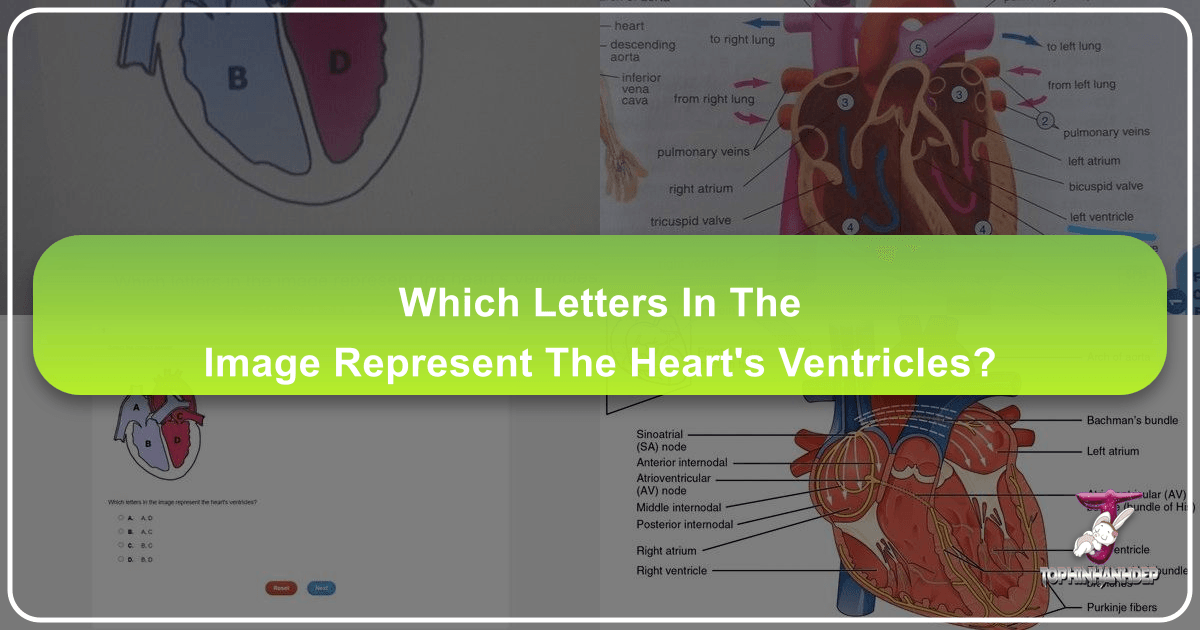
Which Letters in the Image Represent the Heart's Ventricles? Exploring Cardiac Anatomy Through Visuals

Understanding the complex machinery of the human heart is a foundational aspect of biology and medicine. At its core, the heart is a powerful, four-chambered muscular pump, meticulously designed to circulate blood throughout the body. Among these chambers, the ventricles play a paramount role, acting as the primary drivers of blood flow. When presented with anatomical diagrams or visual representations of the heart, the ability to correctly identify these crucial structures, often labeled with letters or numbers, is key to grasping cardiac function. On Tophinhanhdep.com, we recognize the indispensable value of high-quality images, from detailed anatomical illustrations to dynamic photography, in deciphering such intricate biological systems and enhancing learning.
The question “which letters in the image represent the heart’s ventricles?” is more than a simple identification task; it’s an entry point into a deeper understanding of cardiac physiology, blood circulation, and even the electrical activity that drives this vital organ. Through the lens of visual design, photography, and comprehensive image collections, Tophinhanhdep.com provides the resources necessary to not only answer this question accurately but also to explore the broader context of heart health and function.

Visualizing the Heart’s Ventricles: Function and Anatomy
The human heart is divided into four distinct chambers: two atria (upper chambers) and two ventricles (lower chambers). While all chambers are vital, the ventricles bear the significant responsibility of generating the force required to propel blood out of the heart and into the circulatory system. In educational diagrams, identifying these specific chambers is often facilitated by clear labeling, typically using letters or numbers.
The term “ventricle” itself refers to a muscular chamber that actively pumps blood. In humans and other mammals, there are two ventricles: the right ventricle and the left ventricle. These chambers are positioned at the bottom of the heart, forming a somewhat pointed base that often leans towards the left side of the chest. The significance of their muscular walls cannot be overstated; they are considerably more robust than those of the atria because they must exert immense pressure to circulate blood effectively. The left ventricle, in particular, is far more heavily muscled than the right, as it is responsible for pumping oxygen-rich blood to the entire systemic circulation – from the brain down to the toes. The right ventricle, while also powerful, pumps de-oxygenated blood only to the nearby lungs, a much shorter circulatory path.
When encountering an anatomical diagram of the heart on a platform like Tophinhanhdep.com, one might see various letters or numbers pointing to different parts. If, for instance, a question asks what letter ‘D’ represents and the options include ‘aorta, left atrium, left ventricle, right atrium,’ and ‘D’ points to the larger, lower-left chamber, then ‘D’ would represent the left ventricle. Similarly, another letter pointing to the lower-right chamber would denote the right ventricle. The two chambers above them, which receive blood, would be the atria. This clear visual identification is a cornerstone of learning, made accessible through high-resolution images and thoughtfully designed illustrations.

Identifying the Pumping Chambers in Diagrams
The process of pinpointing the ventricles in a visual representation typically involves understanding their relative position and connection points. As the lower chambers, they are visually distinct from the atria. Furthermore, their muscular walls, especially the left ventricle’s, are often depicted as thicker, reflecting their arduous pumping role.
For instance, consider a simplified heart diagram:
- De-oxygenated blood from the body enters the right atrium.
- It then passes into the right ventricle.
- The right ventricle pumps this blood into the pulmonary artery, which carries it to the lungs for oxygenation.
- Freshly oxygenated blood returns from the lungs to the left atrium via the pulmonary veins.
- From the left atrium, it moves into the left ventricle.
- Finally, the left ventricle powerfully pumps this oxygenated blood into the aorta, the body’s main artery, to be distributed throughout the systemic circulation.
In such a flow, any labels directly pointing to the two large, bottom-most chambers that connect to the pulmonary artery and the aorta, respectively, would represent the ventricles. Visual learners benefit immensely from detailed wallpapers and backgrounds on Tophinhanhdep.com that clearly delineate these pathways and structures, allowing for repeated self-testing and reinforcing knowledge. Our AI upscalers can even enhance older diagrams, making historical anatomical drawings clearer and more useful for modern study.

The Structural Intricacies of Ventricular Walls
Beyond just identifying the ventricles, understanding their internal structure adds another layer of appreciation for their function. The walls of these chambers are composed primarily of specialized muscle tissue known as cardiac muscle cells, or cardiomyocytes. These cells are shorter and have smaller diameters compared to skeletal muscle cells but are highly organized, demonstrating striations (alternating dark and light bands) due to the precise arrangement of contractile myofilaments.
Key structural features within the ventricles, often highlighted in detailed anatomical photography available on Tophinhanhdep.com, include:
- Trabeculae Carneae: These are irregular bundles and bands of muscle that ridge the interior surfaces of the ventricles. They contribute to the efficiency of ventricular contraction and help prevent suction effects that might impede blood flow.
- Papillary Muscles: Projecting like small cones or nipples into the ventricular cavities, these muscles are crucial for valve function. Fine strands of tendon, called chordae tendineae, attach from the papillary muscles to the cusps of the atrioventricular valves (tricuspid on the right, mitral on the left). When the ventricles contract, the papillary muscles also contract, pulling on the chordae tendineae to prevent the valve cusps from prolapsing (inverting) back into the atria, ensuring efficient, one-way blood flow.
Cardiac muscle cells themselves possess unique properties, including autorhythmicity, the ability to initiate their own electrical potential at a fixed rate, which then spreads rapidly to trigger contraction. This is a fundamental difference from skeletal muscle. These cells are interconnected by crucial structures called intercalated discs, which are junctions that strongly bind adjacent cells and facilitate the rapid transmission of electrical impulses. These discs contain desmosomes (for strong adhesion) and gap junctions (allowing ion passage), ensuring a synchronized contraction across the ventricular muscle, vital for effective pumping. High-resolution microscopy images and detailed digital art on Tophinhanhdep.com can visually demonstrate these microscopic wonders, bridging the gap between macro-anatomy and cellular physiology.
The Heart’s Electrical System: A Visual Conduction Pathway
The powerful contractions of the ventricles are not random; they are meticulously coordinated by a sophisticated electrical conduction system within the heart. Visual representations, whether diagrams or electrocardiograms (ECGs), are indispensable tools for understanding this intricate electrical symphony. Tophinhanhdep.com offers a wealth of visual aids that simplify complex physiological processes, making them accessible to students and professionals alike.
The heart’s autorhythmicity means it can generate its own electrical impulses. This process begins in specialized conducting cells that form the conduction system, ensuring a precise sequence of contraction that allows the heart to efficiently pump blood.
Tracing the Impulse: From SA Node to Purkinje Fibers
The electrical impulse that ultimately triggers ventricular contraction originates in the sinoatrial (SA) node, often referred to as the heart’s natural pacemaker. Located in the right atrium, the SA node has the highest inherent rate of depolarization, setting the normal rhythm (sinus rhythm) of the heart. From the SA node, the impulse spreads across the atria through specialized internodal pathways and Bachmann’s bundle (to the left atrium), causing the atria to contract and push blood into the ventricles.
Crucially, the impulse then reaches the atrioventricular (AV) node, located in the inferior portion of the right atrium. Here, there’s a critical delay, typically about 100 milliseconds, before the impulse is transmitted further. This pause is vital, allowing the atria to complete their contraction and fully empty their blood into the ventricles before the ventricles themselves begin to contract. Without this delay, the atria and ventricles might contract simultaneously, leading to inefficient blood pumping.
After the AV node delay, the impulse rapidly travels down the atrioventricular bundle (Bundle of His), through the interventricular septum. It then divides into the left and right bundle branches, which extend towards the apex of the heart. The left bundle branch is significantly larger, reflecting the greater muscle mass of the left ventricle it supplies. Finally, these branches connect to a network of specialized conductive fibers called Purkinje fibers, which rapidly spread the impulse throughout the myocardial contractile cells of the ventricles.
The electrical stimulus reaches the apex of the heart first, causing ventricular contraction to begin there and spread upwards towards the base, effectively “squeezing” the blood out of the ventricles into the pulmonary artery and aorta. This entire process, from SA node initiation to full ventricular depolarization, takes approximately 225 milliseconds. Visual aids, such as animated diagrams or step-by-step illustrations found in Tophinhanhdep.com’s collections, are invaluable for visualizing this precise, synchronized electrical flow. Graphic design and digital art tools enable the creation of aesthetic and clear infographics that trace the impulse’s path, making a complex physiological process understandable.
Electrocardiograms (ECGs): A Snapshot of Ventricular Activity
While internal diagrams show the conduction pathway, the electrocardiogram (ECG or EKG) provides a real-time surface recording of the heart’s electrical activity. This non-invasive diagnostic tool relies on the careful placement of electrodes on the body to detect the complex, compound electrical signals generated by the heart. On Tophinhanhdep.com, you can find various illustrations and explainers of ECG tracings, crucial for understanding both normal and abnormal heart function.
A typical normal ECG tracing features several characteristic waves, complexes, and segments, each corresponding to specific electrical events in the cardiac cycle:
- P wave: Represents the depolarization of the atria. This is the electrical signal that initiates atrial contraction.
- QRS complex: This is the largest and most prominent part of the ECG, representing the depolarization of the ventricles. Because the ventricular muscle mass is much larger, it generates a stronger electrical signal. The ventricles begin to contract as the QRS complex reaches its peak. Importantly, the repolarization of the atria also occurs during the QRS complex but is masked by the larger ventricular activity.
- T wave: Represents the repolarization of the ventricles, as the ventricular muscle cells return to their resting electrical state, preparing for the next contraction.
Analyzing the shape, duration, and intervals between these components on an ECG tracing provides critical diagnostic information. For example, an abnormally long PR interval might indicate a delay in conduction from the SA node to the AV node (a “heart block”). An enlarged QRS complex could suggest enlarged ventricles, while an elevated ST segment is a classic sign of an acute myocardial infarction (heart attack).
Tophinhanhdep.com’s image tools, such as AI upscalers, can clarify older or lower-resolution ECG images, making them more amenable to study. Our curated “Image Inspiration & Collections” can provide thematic groupings of normal and abnormal ECGs alongside explanatory diagrams, serving as a comprehensive visual library for medical students and healthcare professionals. Visual design principles employed in creating these educational graphics ensure that even complex abnormalities, such as ventricular fibrillation (a life-threatening uncoordinated beating of the ventricles), are depicted with clarity and accuracy.
Beyond Anatomy: Visualizing Cardiac Health, Disease, and Innovation
The importance of visual content extends beyond basic anatomical identification. Tophinhanhdep.com serves as a hub for images that illustrate various aspects of cardiac health, disease, and the cutting-edge innovations in treatment. From abstract representations of cellular processes to detailed photography of medical devices, visual media transforms complex concepts into understandable narratives.
The Visual Impact of Cardiac Injury and Healing
Unfortunately, the heart is susceptible to various diseases, with heart attacks being one of the most common and devastating. A heart attack, or myocardial infarction (MI), occurs when blood flow to a section of the heart muscle is interrupted, usually due to a blockage (plaque buildup and subsequent clot formation) in the coronary arteries. Without adequate oxygen and nutrients, the affected cardiac muscle cells begin to die.
The visual representation of a heart attack, often depicted in medical illustrations or educational videos, shows arteries narrowing due to plaque (atherosclerosis) and then a clot completely obstructing the flow. The resulting dead tissue, known as an infarct, is starkly different from healthy muscle. The body’s response is often to replace this dead tissue with scar tissue, which is weaker and less functional than the original cardiac muscle. This scar tissue cannot contract or conduct electrical impulses effectively, leading to a weakened heart and potentially irregular heart rhythms.
Diagrams illustrating the progression of atherosclerosis, cross-sections of damaged heart muscle, or even aesthetic abstract interpretations of cellular damage can be found within Tophinhanhdep.com’s diverse image collections. Such visuals are critical for raising public awareness about risk factors (like poor diet, smoking, obesity) and the consequences of neglecting cardiovascular health. Photography that captures the subtle signs of cardiac distress or the visual effects of recovery can also serve as powerful educational tools.
Illustrating Advanced Treatments for Ventricular Dysfunction
In cases of severe heart damage or chronic heart conditions, advanced medical interventions become necessary. Visuals play a crucial role in explaining these complex treatments to patients, educating future medical professionals, and inspiring creative solutions in bioengineering.
- Ventricular Assist Devices (VADs): For patients with weakened ventricles, often due to severe heart failure, a VAD can be implanted. The most common is the Left Ventricular Assist Device (LVAD), a battery-powered pump that helps the left ventricle circulate blood to the body. Tophinhanhdep.com might feature high-resolution photography of these devices, detailed diagrams illustrating their implantation and function, or digital art conceptualizing their benefits. These images visually articulate how mechanical support can restore adequate blood flow and give the heart time to heal or serve as a bridge to transplant.
- Heart Transplants: For end-stage heart failure, a heart transplant may be the only option. This involves surgically replacing the diseased heart with a healthy donor heart. The visual complexity of such a procedure, from surgical diagrams to before-and-after imagery, underscores the monumental achievement of modern medicine.
- Regenerative Medicine: A promising frontier involves regenerating damaged heart tissue, moving beyond scar tissue formation. This often involves using stem cells, which can be stimulated to become new heart cells. Visual concepts, such as illustrations of stem cells differentiating into cardiomyocytes on a scaffold patch, or animations showing new cells integrating into damaged tissue, are vital for conveying the potential of this emerging field. Tophinhanhdep.com’s collection of scientific illustrations and digital art can provide compelling visual narratives for these groundbreaking treatments, transforming abstract scientific principles into tangible, hopeful images.
Tophinhanhdep.com: Your Visual Gateway to Understanding the Heart and Beyond
From answering a specific question like “which letters in the image represent the heart’s ventricles?” to delving into the intricate electrical signals, structural nuances, and groundbreaking treatments of cardiac medicine, Tophinhanhdep.com stands as an invaluable resource. Our platform is dedicated to curating and providing a vast array of high-quality visual content that empowers learning, inspires creativity, and facilitates understanding across a multitude of disciplines.
Whether you are a student grappling with anatomy, a medical professional seeking illustrative resources, a graphic designer creating educational content, or simply someone with an aesthetic appreciation for the complexities of life, Tophinhanhdep.com offers the tools and inspiration you need.
Our Images section, rich with wallpapers, backgrounds, and beautiful photography, includes detailed anatomical renderings that allow for precise identification of structures like the heart’s ventricles. You can find aesthetic and nature-inspired visuals that indirectly reflect the beauty of biological systems, or sad/emotional imagery that might convey the impact of cardiac disease.
In Photography, we emphasize high-resolution and stock photos, ensuring that every detail of a cardiac diagram, microscopic view of a cardiomyocyte, or surgical image is rendered with crystal clarity. Our collections cover various digital photography and editing styles, making it possible to present complex medical information in engaging and accessible formats.
Our Image Tools are designed to optimize and enhance your visual experience. Converters, compressors, and optimizers ensure fast loading and efficient use of images. The AI Upscalers can take a historical medical diagram or an older scan of a heart and transform it into a crisp, high-definition visual, bringing aged educational materials into the modern era. Image-to-text features can even help extract labels from complex diagrams for easier study.
For those involved in Visual Design, Tophinhanhdep.com is a wellspring of inspiration. Graphic design, digital art, and photo manipulation techniques are employed to create stunning and informative visuals. From creative ideas for illustrating blood flow to sophisticated digital art representing cellular processes, our platform supports the creation of impactful educational and inspirational content.
Finally, our Image Inspiration & Collections offer curated photo ideas, mood boards, thematic collections, and trending styles that can inform and elevate any project related to human anatomy or health. Imagine a collection dedicated entirely to “The Pumping Heart,” featuring everything from cross-sections of ventricles to animated representations of ECG waves, all meticulously organized for ease of access and study.
In conclusion, correctly identifying the heart’s ventricles in an image is just the beginning of a fascinating journey into human biology. Tophinhanhdep.com is committed to being your trusted partner in this exploration, providing the visual resources, tools, and inspiration to unlock the secrets of the heart and countless other wonders of the world, one exceptional image at a time.